Найти в Дзене
СфераЖизни

2. Case problems with pictures in topic "Physiology of excitable tissues"

Hi all! I present to your attention case problems with pictures and solutions to them.

Case problem 2.1. The general principle of physiology that structure is a determinant of - and has coevolved with function can be considered at the molecular, cellular, and organ levels. How is this principle illustrated by the binding of messengers to their receptors?

Figure 2.1. Characteristics of receptors binding to messengers. The receptors with high affinity will have more bound messenger at a given messenger concentration (e.g., concentration X). The presence of a competitor will decrease the amount of messenger bound, until at very high concentrations the receptors become saturated with messenger and cannot bind any additional messenger. Note in the illustration that the low-affinity receptor in this case has a slightly different shape in its ligand-binding region compared to the high-affinity receptor, which makes it less able to bind the messenger. Also note the similarity in parts of the shapes of the natural messenger and its competitor. Eric P. Widmaier Human Physiology. The Mechanisms of Body Function, New NY: McGraw-Hill Education. 2019. https://lccn.loc.gov/2017048599

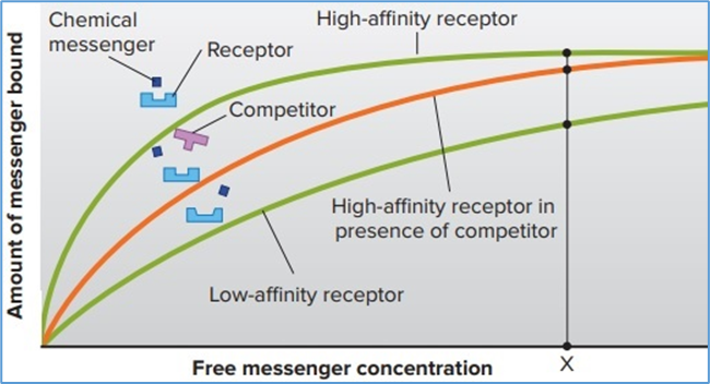
Hint for answer 2.1. The structures of both the messenger and its receptors determine their ability to bind to each other with specificity. It is the binding of a messenger to a receptor that causes the activation (function) of the receptor. In addition, any molecule with a structure that is sufficiently similar to that of the messenger may also bind that receptor; in the case of competitors, this may decrease the function of the messenger-receptor system. The specificity of the messenger-receptor interaction allows each messenger to exert a discrete action. This is the basis of many therapeutic medications that are used to block the deleterious effects of an excess of naturally occurring messengers.

Case problem 2.2. How does the chemical nature of lipid-soluble messengers relate to the general principle of physiology that physiological processes are dictated by the laws of chemistry and physics?

Figure 2.2. Mechanism of action of   lipid-soluble messengers. This figure shows the receptor (simplified in this   view) for these messengers in the nucleus. In some cases, the unbound   receptor is in the cytosol rather than the nucleus, in which case the binding   occurs there, and the activated messenger-receptor complex then moves into   the nucleus. For simplicity, a single messenger is shown binding to a single   receptor. In many cases, however, two messenger receptor complexes must bind   together in order to activate a gene. Eric P. Widmaier Human Physiology. The   Mechanisms of Body Function, New NY: McGraw-Hill Education. 2019. https://lccn.loc.gov/2017048599
Figure 2.2. Mechanism of action of lipid-soluble messengers. This figure shows the receptor (simplified in this view) for these messengers in the nucleus. In some cases, the unbound receptor is in the cytosol rather than the nucleus, in which case the binding occurs there, and the activated messenger-receptor complex then moves into the nucleus. For simplicity, a single messenger is shown binding to a single receptor. In many cases, however, two messenger receptor complexes must bind together in order to activate a gene. Eric P. Widmaier Human Physiology. The Mechanisms of Body Function, New NY: McGraw-Hill Education. 2019. https://lccn.loc.gov/2017048599

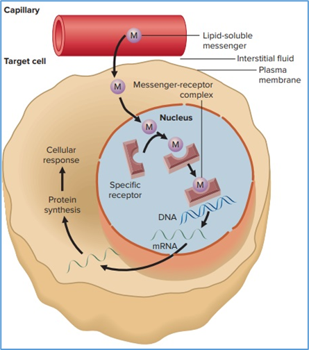
Hint for answer 2.2. The lipid nature of certain messengers makes it possible for them to diffuse through the lipid bilayer of a plasma membrane. Consequently, the receptors for such messengers exist inside the cell. By contrast, hydrophilic messengers cannot penetrate a lipid bilayer, and as a result their receptors are located within plasma membranes with an extracellular component that can detect specific ligands. Therefore, the cellular location of receptors for chemical messengers depends upon the chemical characteristics of the messengers, which, in turn, determines their permeability through cell membranes.

Case problem 2.3. Does a given protein kinase, such as cAMP-dependent protein kinase, only phosphorylate the same proteins in all cells in which the kinase is present?

Figure 2.3. The variety of cellular responses   induced by cAMP is due mainly to the fact that activated cAMP-dependent   protein kinase can phosphorylate many different proteins, activating or   inhibiting them. In this figure, the protein kinase is shown phosphorylating   seven different proteins a microtubular protein, an ATPase, an ion channel, a   protein in the endoplasmic reticulum, a protein involved in stimulating the   transcription of a gene into mRNA, and two enzymes. Eric P. Widmaier Human   Physiology. The Mechanisms of Body Function, New NY: McGraw-Hill Education.   2019. https://lccn.loc.gov/2017048599
Figure 2.3. The variety of cellular responses induced by cAMP is due mainly to the fact that activated cAMP-dependent protein kinase can phosphorylate many different proteins, activating or inhibiting them. In this figure, the protein kinase is shown phosphorylating seven different proteins a microtubular protein, an ATPase, an ion channel, a protein in the endoplasmic reticulum, a protein involved in stimulating the transcription of a gene into mRNA, and two enzymes. Eric P. Widmaier Human Physiology. The Mechanisms of Body Function, New NY: McGraw-Hill Education. 2019. https://lccn.loc.gov/2017048599

Hint for answer 2.3. Not necessarily. In some cases, a kinase may phosphorylate the same protein in many different types of cells. However, many cells also express certain cell-specific proteins that are not found in all tissues, and some of these proteins may be substrates for cAMP-dependent protein kinase. Thus, the proteins that are phosphorylated by a given kinase depend upon the cell type, which makes the cellular response tissue-specific. As an example, in the kidneys, cAMP-dependent protein kinase phosphorylates proteins that insert water channels in cell membranes and thereby decrease urine volume, whereas in heart muscle the same kinase phosphorylates Ca2+ channels that increase the strength of muscle contraction.

Case problem 2.4. In setting up this experiment, 0.15 mole of NaCl was placed in compartment 1, 0.15 mole of KCl was placed in compartment 2, and each compartment has a volume of 1 liter. What is the approximate total solute concentration in each compartment at equilibrium (that is, in panel e)?

Figure 2.4. Generation of a potential across   a membrane due to diffusion of K+ through K+ channels   (red). Arrows represent ion movements; arrow length represents the magnitude   of the flux. Eric P. Widmaier Human Physiology. The Mechanisms of Body   Function, New NY: McGraw-Hill Education. 2019.   https://lccn.loc.gov/2017048599
Figure 2.4. Generation of a potential across a membrane due to diffusion of K+ through K+ channels (red). Arrows represent ion movements; arrow length represents the magnitude of the flux. Eric P. Widmaier Human Physiology. The Mechanisms of Body Function, New NY: McGraw-Hill Education. 2019. https://lccn.loc.gov/2017048599

Hint for answer 2.4. NaCl and KCl ionize in solution virtually completely, so initially each compartment would have a total solute concentration of approximately 0.3 osmols per liter (see Chapter 4 to review the difference between moles and osmols). Because an insignificant number of potassium ions actually move in establishing the equilibrium potential, the final solute concentrations of the compartments would not be significantly different.

Case problem 2.5. In this hypothetical system, what would the concentrations of each ion be at equilibrium (panel e) if open channels for both Na+ and K+ were present?

Figure 2.5. Generation of a potential across   a membrane due to diffusion of Na+ through Na+ channels   (blue). Arrows represent ion movements, arrow length indicates the magnitude   of the flux. So, few sodium ions cross the membrane that ion concentrations   do not change significantly from step (a) to step (e). See the text for a   more complete explanation. Eric P. Widmaier Human Physiology. The Mechanisms   of Body Function, New NY: McGraw-Hill Education. 2019.   https://lccn.loc.gov/2017048599
Figure 2.5. Generation of a potential across a membrane due to diffusion of Na+ through Na+ channels (blue). Arrows represent ion movements, arrow length indicates the magnitude of the flux. So, few sodium ions cross the membrane that ion concentrations do not change significantly from step (a) to step (e). See the text for a more complete explanation. Eric P. Widmaier Human Physiology. The Mechanisms of Body Function, New NY: McGraw-Hill Education. 2019. https://lccn.loc.gov/2017048599

Hint for answer 2.5. Na+ and K+ would move down their concentration gradients in opposite directions, each canceling the charge carried by the other. Thus, at equilibrium, there would be no membrane potential and both compartments would have 0.15 M Cl−, 0.075 M Na+, and 0.075 M K+.

Case problem 2.6. Would decreasing a neuron’s intracellular fluid [K+] by 1 mM have the same effect on resting membrane potential as raising the extracellular fluid [K+] by 1 mM?

Figure 2.6. Forces influencing sodium and   potassium ions at the resting membrane potential (Vm). (a) At a resting   membrane potential of −70 mV, both the concentration and electrical gradients   favor inward movement of Na+, whereas the K+    concentration and electrical gradients are in opposite directions. (b) The   greater permeability of K+ maintains the resting membrane potential at a   value near EK. Eric P. Widmaier Human Physiology. The Mechanisms of Body   Function, New NY: McGraw-Hill Education. 2019. https://lccn.loc.gov/2017048599
Figure 2.6. Forces influencing sodium and potassium ions at the resting membrane potential (Vm). (a) At a resting membrane potential of −70 mV, both the concentration and electrical gradients favor inward movement of Na+, whereas the K+ concentration and electrical gradients are in opposite directions. (b) The greater permeability of K+ maintains the resting membrane potential at a value near EK. Eric P. Widmaier Human Physiology. The Mechanisms of Body Function, New NY: McGraw-Hill Education. 2019. https://lccn.loc.gov/2017048599

Hint for answer 2.6.Changing the ECF [K+] has a greater effect on EK (and thus the resting membrane potential). This is because the ratio of external to internal K+ is changed more when ECF concentration goes from 5 to 6 mM (a 20% increase) than when ICF concentration is decreased from 150 to 149 mM (a 0.7% decrease). You can confirm this with the Nernst equation. Inserting typical values, when [Kout] = 5 mM and [Kin] = 150 mM, the calculated value of EK = −90.1 mV. If you change [Kin] to 149 mM, the calculated value of EK = −89.9 mV, which is not very different. By comparison, changing [Kout] to 6 mM causes a greater change, with the resulting EK = −85.3 mV.

Case problem 2.7. If the ligand-gated ion channel allowed only K+ movement, how would this figure be different?

Figure 2.7. Depolarisation and graded potential caused by a chemical stimulus. Inward positive current through ligand-gated cation channels depolarizes a region of the membrane, and local currents spread the depolarization to adjacent regions. Eric P. Widmaier Human Physiology. The Mechanisms of Body Function, New NY: McGraw-Hill Education. 2019. https://lccn.loc.gov/2017048599

Hint for answer 2.7. K+ would exit from the cell and make the inside of the cell more negative in the area of the channel, and thus positive current would flow toward the channel’s location on the inside of the cell and away from the channel on the outside.

Case problem 2.8. If extracellular [Na+] is elevated, how would the resting potential and action potential of a neuron change?

Figure 2.8. The changes in (a) membrane potential and (b) relative membrane permeability (P) to sodium and potassium ions during an action potential. Eric P. Widmaier Human Physiology. The Mechanisms of Body Function, New NY: McGraw-Hill Education. 2019. https://lccn.loc.gov/2017048599

Hint for answer 2.8. The value of the resting potential would change very little because the permeability of resting membranes to Na+ is very low. However, during an action potential, the membrane voltage would rise more steeply and reach a more positive value due to the larger electrochemical gradient for Na+ entry through open voltage-gated ion channels.

Case problem 2.9. Striking the ulnar nerve in your elbow against a hard surface initiates action potentials near the midpoint of sensory and motor axons traveling in that nerve. In which direction will those action potentials propagate?

Figure 2.9. One-way propagation of an action potential. For simplicity, potentials are shown only on the upper membrane, local currents are shown only on the inside of the membrane, and repolarizing currents are not shown. (a) Local current from the opening of ligand-gated ion channels in the cell body and dendrites causes an action potential to be initiated in region 1, and local current depolarizes region 2. (b) Action potential in region 2 generates local currents; region 3 is depolarized toward threshold, but region 1 is refractory. (c) Action potential in region 3 generates local currents, but region 2 is refractory. Eric P. Widmaier Human Physiology. The Mechanisms of Body Function, New NY: McGraw-Hill Education. 2019. https://lccn.loc.gov/2017048599

Hint for answer 2.9. In all of the affected neurons, action potentials will propagate in both directions from the elbow - up the arm toward the spinal cord and down the arm toward the hand. Action potentials traveling upward along afferent pathways will continue through synapses into the CNS to be perceived as pain, tingling, vibration, and other sensations of the lower arm. In contrast, action potentials traveling backward up motor axons will die out once they reach the cell bodies because synapses found there are “one way” in the opposite direction.

Case problem 2.10. A general principle of physiology states that homeostasis is essential for health and survival. In what ways might the presence of myelin contribute to homeostasis?

Figure 2.10. Myelinisation and saltatory conduction of action potentials. K+ channels are not depicted (they are located primarily at the myelin/node junctions and help to repolarise the neuron). As described in Figure 6.15, positive charges move away from the site of depolarisation on the inside of the cell, and toward it on the outside. Eric P. Widmaier Human Physiology. The Mechanisms of Body Function, New NY: McGraw-Hill Education. 2019. https://lccn.loc.gov/2017048599

Hint for answer 2.10. Myelin increases conduction speed along an axon, which is important for rapid signaling and reflexes. As just one common example, fast motor reflexes may help prevent injury by removing a part of the body (such as your hand) from danger, such as a sharp or burning object. If your hand did not quickly pull away from such harmful objects, much more severe injury would occur. Myelin also decreases the metabolic cost of sending electrical signals along axons, thereby saving energy for other homeostatic processes.

Case problem 2.11. Assuming a typical EPSP of 0.5 mV, approximately how many simultaneous EPSPs would be required to bring a typical neuron to threshold?

Figure 2.11. Excitatory postsynaptic potential   (EPSP). Stimulation of the presynaptic neuron is marked by the green arrow.   (Drawn larger than normal: typical EPSP = 0.5 mV). Eric P.   Widmaier Human Physiology. The Mechanisms of Body Function, New NY:   McGraw-Hill Education. 2019. https://lccn.loc.gov/2017048599
Figure 2.11. Excitatory postsynaptic potential (EPSP). Stimulation of the presynaptic neuron is marked by the green arrow. (Drawn larger than normal: typical EPSP = 0.5 mV). Eric P. Widmaier Human Physiology. The Mechanisms of Body Function, New NY: McGraw-Hill Education. 2019. https://lccn.loc.gov/2017048599

Hint for answer 2.11. In a typical neuron, the threshold potential is about 15 mV more positive than the resting membrane potential, so it would take about 30 simultaneous EPSPs of 0.5 mV to reach threshold.

Case problem 2.12. How might the traces in part 5 be different if the excitatory synapse (A) was much closer to the axon hillock than the inhibitory synapse (C)?

Figure 2.12. Interaction of EPSPs and IPSPs at the postsynaptic neuron. Presynaptic neurons (A–C) were stimulated at times indicated by the arrows, and the resulting membrane potential was recorded in the postsynaptic cell by a recording microelectrode. Eric P. Widmaier Human Physiology. The Mechanisms of Body Function, New NY: McGraw-Hill Education. 2019. https://lccn.loc.gov/2017048599

Hint for answer 2.12. The greater the distance between the synapse and the axon hillock (the location of the electrode), the greater the decrement of a graded potential. Therefore, if synapse A were closer to the axon hillock than synapse C, summing the two would most likely result in a small depolarizing potential. The farther from the hillock synapse C is, the more closely the depolarization would come to resemble the trace occurring in response to synapse A firing alone.

Case problem 2.13. How would this afferent pathway be affected by exposing this entire neuron to a drug that blocks voltage-gated Ca2+ channels? (Recall from Sections B and C in Chapter 6 which ions are involved in different aspects of neuronal signaling).

Figure 2.13. Stimulation of an afferent neuron with a receptor ending. Electrodes   measure graded potentials and action potentials at various points in response   to different stimulus intensities. Action potentials arise at the first node   of Ranvier in response to a suprathreshold stimulus, and the action potential   frequency and neurotransmitter release increase as the stimulus and receptor   potential become larger. Eric P. Widmaier   Human Physiology. The Mechanisms of Body Function, New NY: McGraw-Hill   Education. 2019. https://lccn.loc.gov/2017048599
Figure 2.13. Stimulation of an afferent neuron with a receptor ending. Electrodes measure graded potentials and action potentials at various points in response to different stimulus intensities. Action potentials arise at the first node of Ranvier in response to a suprathreshold stimulus, and the action potential frequency and neurotransmitter release increase as the stimulus and receptor potential become larger. Eric P. Widmaier Human Physiology. The Mechanisms of Body Function, New NY: McGraw-Hill Education. 2019. https://lccn.loc.gov/2017048599

Hint for answer 2.13. Receptor potentials would not be affected because they are not mediated by voltage-gated ion channels. Action potential propagation to the central nervous system would also be normal because it depends only on voltage-gated Na+ and K+ channels. The drug would inhibit neurotransmitter release from the central axon terminal, however, because vesicle exocytosis requires Ca2+ entry through voltage-gated ion channels.

Thank you for attention! Good luck in your studies! :-)