Good afternoon!
Case problem 9.1. A patient suffering from cholera, a disease that causes severe diarrhea, produces 12 liters of watery feces in one day, despite ingesting only 1.5 liters of water and 500 grams of solid food. Where does the extra water come from?
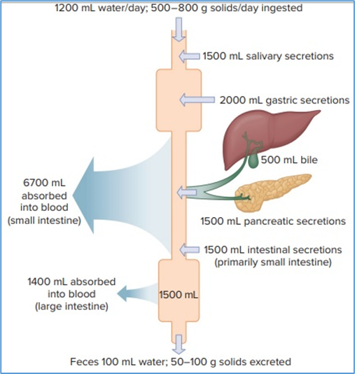
Hint for answer 9.1. The large quantities of fluid that exit the body in diarrheal illnesses are not simply due to ingested fluids and foods not being absorbed. Large volumes of fluid entering the tract across the intestinal wall come from the body’s interstitial fluid and plasma compartments. Loss of such large quantities of fluid from the body (dehydration) is what can make these disorders fatal. Patients must be supplied with large quantities of ingested or intravenous fluids in order to survive. The most common finding is an abnormally high production of gastric (hydrochloric) acid due to gastrin stimulation of the parietal cell of the stomach. This high acidity can cause injury to the duodenum because the pancreas cannot produce sufficient quantities of HCO3− to neutralise it. The low pH in the duodenum can also inactivate pancreatic enzymes, which can ultimately lead to diarrhea due to unabsorbed nutrients and increased fat in the stool. The spectrum of findings in a patient with a gastrinoma is called the Zollinger-Ellison syndrome.
Case problem 9.2. Referring to parts (b) and (c), what are some of the consequences of aspiration?
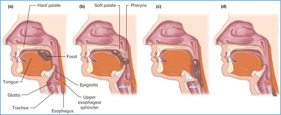
Hint for answer 9.2. Aspiration of food during swallowing can lead to occlusion (blockage) of the airways, which can result in a disruption of oxygen delivery and carbon dioxide removal from the pulmonary system. Aspiration of stomach contents can lead to severe lung damage primarily due to the low pH of the material.
Case problem 9.3. Why doesn’t the high concentration of H+ in the stomach lumen destroy the lining of the stomach wall? (What secretory product protects the stomach?)
Hint for answer 9.3. Mucus secreted by the cells in the gastric gland creates a protective coating and traps HCO3−. This gastric mucosal barrier protects the stomach from the luminal acidity.
Case problem 9.4. What would happen to gastrin secretion in a patient taking a medication that blocks the binding of histamine to its receptor on the parietal cell?
Hint for answer 9.4. A decrease in histamine action would result in a decrease in acid secretion and an increase in the pH of the material in the lumen of the stomach. This would decrease the H+-induced inhibition of gastrin secretion; consequently, gastrin secretion would increase. Because a large part of the effect of gastrin on acid secretion is by stimulating histamine release, the parietal cell acid secretion would still be decreased. This is why histamine-receptor blockers (called H2 blockers) are effective in increasing stomach pH and alleviating the symptoms of gastroesophageal reflux (heartburn) described earlier in this chapter.
Case problem 9.5. What might occur if a person whose stomach has been removed eats a large meal?
Hint for answer 9.5. A person whose stomach has been removed because of disease (e.g., cancer) must eat more frequent small meals instead of the usual three large meals per day. A large meal in the absence of the controlled emptying by the stomach could rapidly enter the intestine, producing a hypertonic solution. This hypertonic solution could cause enough water to flow (by osmosis) into the intestine from the blood to lower the blood volume and produce circulatory complications. The large distension of the intestine by the entering fluid can also trigger vomiting in such individuals. All of these symptoms produced by the rapid entry of large quantities of ingested material into the small intestine are known as the dumping syndrome.
Case problem 9.6. Do you recall learning about a brush border in any other body structure? (Hint: Think about the functional units of the kidneys).
Hint for answer 9.6. A brush border is also found along the luminal surface of the proximal tubules of the renal nephrons. Like the intestinal brush border, that of the proximal tubules is an adaptation that increases surface area and allows for increased transport of solutes across the epithelium.
Case problem 9.7. In addition to the hepatic portal vein, can you name another portal vein system and explain the meaning of the term portal?
Hint for answer 9.7. A portal vein carries blood from one capillary bed to another capillary bed. The hypothalamo-pituitary portal veins carry hypophysiotropic hormones from the capillaries of the median eminence to the anterior pituitary gland where they stimulate or inhibit the release of pituitary gland hormones.
Case problem 9.8. How do the digestion and absorption of fats illustrate the general principle of physiology that controlled exchange of materials occurs between compartments and across cellular membranes? What types of compartments and membranes are involved, and how are the processes controlled?
Hint for answer 9.8. Exchange of materials occurs across an epithelium from the lumen of the intestine into the central lacteal (the lymph). This process is controlled by the enzymatic breakdown of triglycerides in fat droplets, and the temporary storage of the breakdown products in micelles. Fatty acids and monoglycerides are slowly released from micelles as these products diffuse into epithelial cells. Diffusion is maintained by synthesising new triglycerides in the epithelial cells from the absorbed fatty acids and monoglycerides. Control, therefore, occurs at multiple sites from initial digestion to transepithelial movement of digestion products.
Case problem 9.9. A general principle of physiology is that the functions of organ systems are coordinated with each other. Give several examples of how the functions of the nervous and digestive systems are coordinated.
Hint for answer 9.9. Reflexes mediated by signals from the nervous system to the walls of the stomach and intestine trigger activation of smooth muscle and secretory glands in these organs. In addition, neural input from the autonomic nervous system helps regulate acid production in the stomach and the rate of gastric emptying, as well as the motility of the small intestine.
Good luck in your studies!