Good afternoon!
Case problem 7.1. What consequences would result if inflammation caused a buildup of fluid in the alveoli and interstitial spaces?
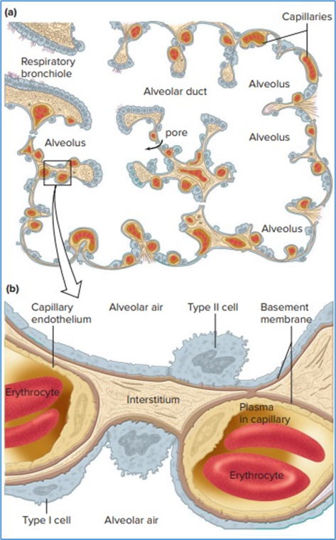
Hint for answer 7.1. The rate of diffusion of gases between the air and the capillaries may be decreased due to the increased resistance to diffusion.
Case problem 7.2. How can a collapsed lung be re-expanded in a patient with a pneumothorax? (Hint: What changes in Pip and Ptp would be needed to re-expand the lung?)
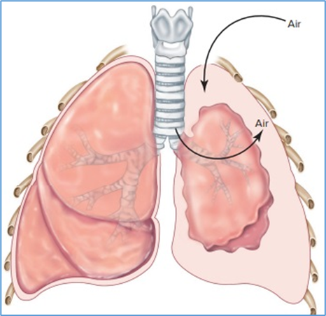
Hint for answer 7.2. A tube is placed through the chest wall into the now enlarged pleural space. (The original hole causing the pneumothorax would need to be repaired first.) Suction is then applied to the chest tube. The negative pressure decreases Pip below Patm and thereby increases Ptp, which results in re-expansion of the lung.
Case problem 7.3. How do the changes in Ptp between each step (–) explain whether the volume of the lung is increasing or decreasing?
Hint for answer 7.3.
Case problem 7.4. Premature infants with inadequate surfactant have decreased lung compliance (respiratory distress syndrome of the newborn). If surfactant is not available to administer for therapy, what would you suggest could be done to inflate the lung?
Hint for answer 7.4. Anything that increases Ptp during inspiration will, theoretically, increase lung volume. This can be done with positive airway pressure generated by mechanical ventilation, which will increase Palv. This approach can work but also increases the risk of pneumothorax by inducing air leaks from the lung into the intrapleural space.
Case problem 7.5. What would be the effect of breathing through a plastic tube with a length of 20 cm and diameter of 4 cm? (Hint: Use the formula for the volume of a perfect cylinder.)
Hint for answer 7.5. The anatomical dead space would be increased by about 251 mL (or 251 cm3). (The volume of the tube can be approximated as that of a perfect cylinder [π r2h = 3.1416 × 22 × 20].) This large increase in anatomical dead space would decrease alveolar ventilation, and tidal volume would have to be increased in compensation. (There would also be an increase in airway resistance.)
Case problem 7.6. How does this figure illustrate the general principle of physiology described in Chapter 1 that physiological processes require the transfer and balance of matter and energy?
Hint for answer 7.6. The cells require oxygen for cellular respiration and, in turn, produce carbon dioxide as a toxic metabolic waste product. To support the net uptake of oxygen and net removal of carbon dioxide, oxygen must be transferred from the atmosphere to all of the cells and organs of the body while carbon dioxide must be transferred from the cells to the atmosphere. This requires a highly efficient transport process that involves diffusion of oxygen and carbon dioxide in opposite directions in the lungs and the cells, and bulk flow of blood carrying oxygen and carbon dioxide around the circulatory system from the lungs to the cells and then back to the lungs. These processes result in a net gain of oxygen (250 ml/per min at rest) from the atmosphere for consumption in the cells, and the net loss of carbon dioxide (200 ml/min at rest) from the cells to the atmosphere.
Case problem 7.7. What is the effect of strenuous exercise on PO2 at the end of a capillary in a normal region of the lung? In a region of the lung with diffusion limitation due to disease?
Hint for answer 7.7. The increase in cardiac output with exercise greatly increases pulmonary blood flow and decreases the amount of time erythrocytes are exposed to increased oxygen from the alveoli. In a normal region of the lung, there is a large safety factor such that a large increase in blood flow still allows normal oxygen uptake. However, even small increases in the rate of capillary blood flow in a diseased portion of the lung will decrease oxygen uptake due to a loss of this safety factor.
Case problem 7.8. How would you treat carbon monoxide poisoning? Look back at Equation 13.8 and realize that CO can be displaced from hemoglobin binding if the dissolved O2 concentration (PO2) is increased enough. That is, CO and O2 compete for the same binding sites on hemoglobin albeit with CO having a 210 times higher affinity.
Hint for answer 7.8. The most important treatment is to displace CO from hemoglobin with O2. Although CO binds to hemoglobin more avidly than O2, it can dissociate from the hemoglobin molecule if forced off by increasing dissolved O2. This is initially done by giving the patient 100% O2 to breathe to increase the inspired and arterial PO2 as high as possible while ensuring that alveolar ventilation is maintained to rid the body of the carbon monoxide. If the CO poisoning is severe, the patient must be transported to a facility that can give hyperbaric O2 therapy. By increasing the barometric pressure, the inspired and arterial PO2 can be dramatically increased. Ultimately, the success of the therapy depends on the length of time the patient has been exposed to high CO, the magnitude of the CO poisoning, and the rapidity of the reduction in CO binding to hemoglobin in order to restore O2 delivery to the tissues.
Case problem 7.9. Several decades ago, removal of the carotid bodies was tried as a treatment for asthma. It was thought that it would reduce shortness of breath and airway hyperreactivity. What would be the effect of bilateral carotid body removal on someone taking a trip to the top of a mountain (an altitude of 3000 meters)?
Hint for answer 7.9. The ventilatory response to the hypoxia of altitude would be greatly diminished, and it is likely that the person would be extremely hypoxemic as a result. Carotid body removal did not help in the treatment of asthma, and this approach was abandoned.
Case problem 7.10. How does this figure illustrate the general principle of physiology described in Chapter 1 that homeostasis is essential for health and survival?
Hint for answer 7.10. An adequate supply of oxygen to all cells is required for normal organ function, and maintenance of oxygen delivery in the face of decreased oxygen uptake in the lung is an important homeostatic reflex. The most common cause of a decrease in the inspired PO2 is temporary or permanent habitation at altitude, where the atmospheric pressure and therefore the PO2 of the air is lower than at sea level. Without compensation for the lower inspired PO2, arterial blood PO2 could decrease to life-threatening levels. All homeostatic processes in the body depend on a continual input of energy derived from heat or ATP; synthesis of ATP requires oxygen. The arterial chemoreceptors can detect a decrease in arterial PO2 that results from ascent to high altitude and reflexively increase alveolar ventilation to enhance oxygen uptake from the air into the pulmonary capillaries for delivery to the rest of the body. The inability to adequately increase alveolar ventilation at altitude can result in harmful consequences leading to organ damage and even death.
Case problem 7.11. The existence of chemoreceptors in the pulmonary artery has been suggested. Hypothesise a function for peripheral chemoreceptors located on and sensing the PO2 and PCO2 of the blood in the pulmonary artery.
Hint for answer 7.11. These receptors may facilitate the increase in alveolar ventilation that occurs during exercise because pulmonary artery PO2 will decrease and pulmonary artery PCO2 will increase. This would match the increase in tissue metabolism to the increase in alveolar ventilation.
Good luck in your studies!