Good afternoon!
Case problem 4.1. What is the advantage of packaging peptide hormones in secretory vesicles?
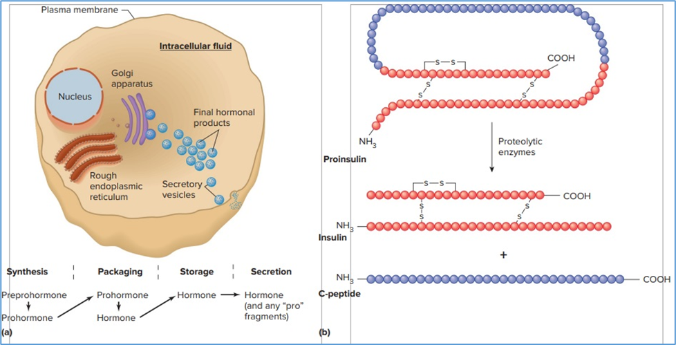
Hint for answer 4.1. By storing large amounts of hormone in an endocrine cell, the plasma concentration of the hormone can be increased within seconds when the cell is stimulated. Such rapid responses may be critical for an appropriate response to a challenge to homeostasis. Packaging peptides in this way also prevents intracellular degradation.
Case problem 4.2. A patient is observed to have symptoms that are consistent with increased concentrations of epinephrine in the blood, including a rapid heart rate, anxiety, and elevated fatty acid concentrations. However, the circulating epinephrine concentrations are measured and found to be in the normal range. What might explain this?
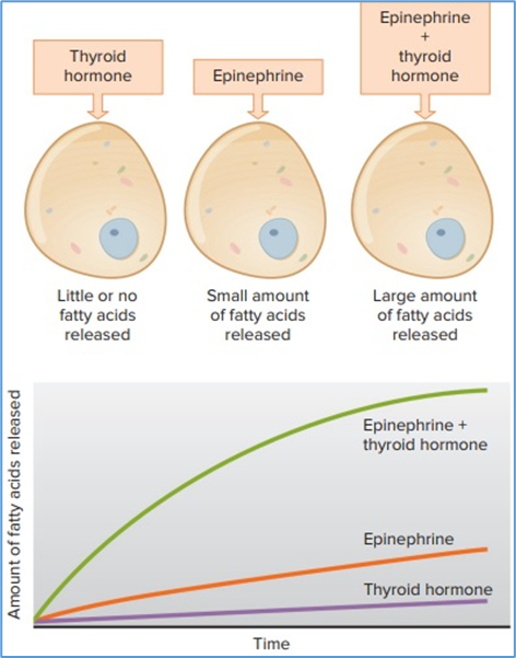
Hint for answer 4.2. One explanation for this patient’s symptoms may be that his or her circulating concentration of thyroid hormone was increased. This might occur if the person’s thyroid was overstimulated due, for example, to thyroid disease. The increased concentration of thyroid hormone would cause an even greater potentiation of the actions of epinephrine, making it appear as if the patient had excess concentrations of epinephrine.
Case problem 4.3. List the several ways this figure illustrates the general principle of physiology described in Chapter 1 that information flow between cells, tissues, and organs is an essential feature of homeostasis and allows for integration of physiological processes.
Hint for answer 4.3. This figure demonstrates how the central nervous system (brain and spinal cord) is the source of afferent information flow that controls many hormonal systems that, in turn, regulate numerous homeostatic processes. For example, the central nervous system is involved in the control of (1) circulatory and metabolic function via release of epinephrine from the adrenal medulla; (2) gastrointestinal function via input from autonomic ganglia to endocrine cells in the intestine; and (3) growth, reproduction, ion and water homeostasis, immune function, and other homeostatic processes via the release of hormones from the anterior and posterior pituitary. This allows a consistent response throughout the body to threats to homeostasis sent by afferent information from throughout the body to the central nervous system, where the information is interpreted and an appropriate response is generated.
Case problem 4.4. Why does it take only very small quantities of hypophysiotropic hormones to achieve concentrations that are effective in regulating anterior pituitary gland hormone secretion?
Hint for answer 4.4. Because the volume of blood into which the hypophysiotropic hormones are secreted is far less than would be the case if they were secreted into the general circulation of the body, the absolute amount of hormone required to achieve a given concentration is much less. This means that the cells of the hypothalamus need only synthesize a tiny amount of hypophysiotropic hormone to reach concentrations in the portal blood vessels that are physiologically active (i.e., can activate receptors on pituitary cells). This allows for the tight control of the anterior pituitary gland by a very small number of discrete neurons within the hypothalamus.
Case problem 4.5. What is the benefit of storing iodinated thyroglobulin in the colloid?
Hint for answer 4.5. Iodine is not widely found in foods; in the absence of iodized salt, an acute or chronic deficiency in dietary iodine is possible. The colloid permits a long-term store of iodinated thyroglobulin that can be used during times when dietary iodine intake is reduced or absent.
Case problem 4.6. What hormonal changes in this pathway would be expected if a patient developed a benign tumor of the left adrenal cortex that secreted extremely large amounts of cortisol in the absence of external stimulation? What might happen to the right adrenal gland?
Hint for answer 4.6. Plasma cortisol concentrations would increase. This would result in decreased ACTH concentrations in the systemic blood, and CRH concentrations in the portal vein blood, due to increased negative feedback at the pituitary gland and hypothalamus, respectively. The right adrenal gland would shrink in size (atrophy) as a consequence of the decreased ACTH concentrations (decreased “trophic” stimulation of the adrenal cortex).
Case problem 4.7. What might happen to plasma concentrations of GH in a person who was intravenously infused with a solution containing a high concentration of glucose, such that his plasma glucose concentrations were significantly increased?
Hint for answer 4.7. Note from the figure that a decrease in plasma glucose concentrations results in an increase in growth hormone concentrations. This makes sense, because one of the metabolic actions of growth hormone is to increase the concentrations of glucose in the blood. By the same reasoning, an increase in the concentration of glucose in the blood due to any cause, including an intravenous infusion as described here, would be expected to decrease circulating concentrations of growth hormone.
Case problem 4.8. Explain how this figure illustrates the general principle of physiology outlined in Chapter 1 that the functions of organ systems are coordinated with each other.
Hint for answer 4.8. The response to hypocalcemia is an excellent example of how the responses of different organ systems function together to restore homeostasis. In this case, the sensor for decreased Ca2+ in the plasma is located in cells of the parathyroid gland. The decrease in Ca2+ increases the synthesis and release of parathyroid hormone (PTH) from these cells. PTH, in turn, coordinates a response of several organ systems to restore plasma Ca2+ to normal. This includes direct effects of PTH on bone to increase resorption (reclamation) of Ca2+ from its storage sites, and on the kidneys to minimize the loss of Ca2+ in the urine as well as to stimulate the production of 1,25-(OH)2D (the active end product of the vitamin D pathway). 1,25-(OH)2D then stimulates an increase in Ca2+ absorption from the small intestine. In this way, an increase in net Ca2+ retention to restore plasma Ca2+ to normal is coordinated by the combined actions of the endocrine, digestive, musculoskeletal, and urinary systems.
Case problem 4.9. Sarcoidosis is a disease that affects a variety of organs (usually the lungs). It is characterized by the development of nodules of inflamed tissue known as granulomas. These granulomas can express significant 1-hydroxylase activity that is not controlled by parathyroid hormone. What will happen to plasma Ca2+ and parathyroid hormone concentrations under these circumstances?
Hint for answer 4.9. The 1-hydroxylase activity will stimulate the conversion of 25-OH D to 1,25-(OH)2D in the granulomas themselves; the 1,25-(OH)2D will then diffuse out of the granuloma cells and enter the plasma, leading to increased Ca2+ absorption in the gastrointestinal tract. This will increase plasma Ca2+, which in turn will suppress parathyroid hormone production; consequently, plasma parathyroid hormone concentrations will decrease. This is a form of secondary hypoparathyroidism.
Good luck in your studies!