Good afternoon, dear students! Posted here case problems with pictures in topic "Physiology of the central nervous system
Case problem 3.1. Describe how the central and peripheral nervous systems illustrate the general principle of physiology that information flow between cells, tissues, and organs is an essential feature of homeostasis and allows for integration of physiological processes.
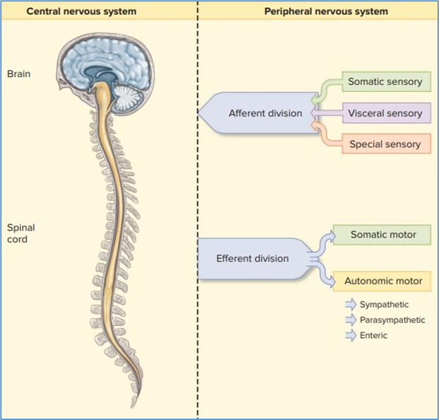
Hint for answer 3.1. Information in the form of electrical signals moves in both directions between the CNS and PNS. In this way, the CNS can be informed of changes in the periphery, such as sensory inputs. In turn, information flow from the CNS to the periphery can direct motor functions that provide an appropriate response to sensory inputs from the PNS. The coordination of sensory and motor inputs and outputs is a key way in which homeostasis is achieved and maintained in the body.
Case problem 3.2. How would the effects differ between a medication that blocks muscarinic acetylcholine receptors and one that blocks nicotinic acetylcholine receptors?
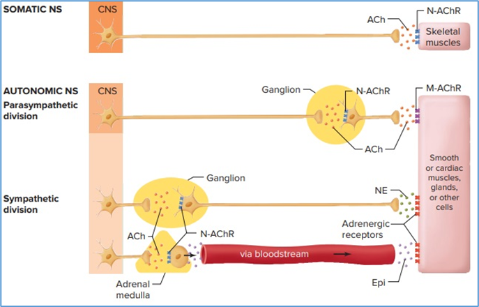
Hint for answer 3.2. The muscarinic receptor blocker would only inhibit parasympathetic pathways, where acetylcholine released from postganglionic neurons binds to muscarinic receptors on target organs. This would reduce the ability to stimulate “rest-or-digest” processes and leave the sympathetic “fight-or-flight” response intact. On the other hand, a nicotinic acetylcholine receptor blocker would inhibit all autonomic control of target organs because those receptors are found at the ganglion in both parasympathetic and sympathetic pathways
Case problem 3.3. Many spinal cord interneurons release the neurotransmitter glycine, which opens chloride ion channels on postsynaptic cell membranes. Given that the plant-derived chemical strychnine blocks glycine receptors, predict the symptoms of strychnine poisoning.
Hint for answer 3.3. Recall that when chloride ion channels are opened, a neuron is inhibited from depolarizing to threshold. Thus, the neurons of the spinal cord that release glycine are inhibitory interneurons. By specifically blocking glycine receptors, strychnine shifts the balance of inputs to motor neurons in favor of excitatory interneurons, resulting in excessive excitation. Poisoning victims experience excessive and uncontrollable muscle contractions body-wide; when the respiratory muscles are affected, asphyxiation can occur. These symptoms are similar to those observed in the disease state tetanus, which is described in the Clinical Case Study at the end of this chapter.
Case problem 3.4. Based on this figure, hypothesize what might happen if you could suddenly stimulate gamma motor neurons to leg flexor muscles in a resting subject.
Hint for answer 3.4. Stimulation of gamma motor neurons to leg flexor muscles would stretch muscle-spindle receptors in those muscles. That would trigger a monosynaptic reflex that would cause contraction of the flexor muscles and, through an interneuron, the extensor muscles would be inhibited. As a result, there would be a reflexive bending of the leg - the opposite of what occurs in the typical knee-jerk reflex.
Case problem 3.5. Which of these conditions would result in the greatest action potential frequency in afferent neurons from muscle-spindle receptors?
Hint for answer 3.5. Although the contracting muscle results in the greatest stretch of the tendon, the muscle itself (and consequently the intrafusal fibers) are stretched the most under passive stretch conditions. Action potentials from muscle-spindle receptors would therefore have the greatest frequency during passive stretch.
Case problem 3.6. While crawling across a floor, a child accidentally places her right hand onto a piece of broken glass. How will the flexor muscles of her left arm respond?
Hint for answer 3.6. When crawling, the crossed-extensor reflex will occur for the arms just like it does in the legs during walking. Afferent pain pathways will stimulate flexor muscles and inhibit extensor muscles in the right arm, while stimulating extensor muscles and inhibiting flexor muscles in the left arm. This withdraws the right hand from the painful stimulus while the left arm straightens to bear the child’s weight.
Case problem 3.7. What structural features of the primary motor cortex somatotopic map reflect the general principle of physiology that structure is a determinant of - and has coevolved with - function?
Hint for answer 3.7. Different regions of the primary motor cortex have evolved different numbers of neurons associated with the specific features of the movements of particular body parts. In this way, the structural organisation of the primary motor cortex is correlated with the functional ability of different body parts. An example is the fine motor control necessary for the movement of fingers while playing a piano; such movements require many more motor neurons than does the ability to move one’s toes.
Case problem 3.8. If a blood clot blocked a cerebral blood vessel supplying a small region of the right cerebral cortex just in front of the central sulcus in the deep groove between the hemispheres, what symptoms might result?
Hint for answer 3.8. When a region of the brain is deprived of oxygen and nutrients for even a short time, it often results in a stroke - neuronal cell death Because the right primary motor cortex was damaged in this case, the patient would have impaired motor function on the left side of the body. Given the midline location of the lesion, the leg would be most affected.
Case problem 3.9. The effect of gravity on stable posture reflects the general principle of physiology that physiological processes are dictated by the laws of chemistry and physics. List other ways you can imagine in which gravity influences physiological functions, including but not limited to motor function.
Hint for answer 3.9. Gravity not only influences posture and balance but also places constraints on many types of motor behaviors, such as jumping or even walking. Simply lifting one’s leg up to take a step requires energy to overcome gravity and to maintain a stable posture and gait. In addition, gravity influences the movement of fluids in the body, such as the flow of blood up to one’s head while standing.
Case problem 3.10. How might the posture shown in part (b) influence contractions of this individual’s shoulder muscles?
Hint for answer 3.10. To stand on the right foot, the hip extensors on the right side are activated while the hip flexors on the left side are activated. This is similar to what occurs when a walking person lifts the left leg and pushes forward with the right foot. In adults, spinal cord interneurons form locomotor pattern generators that connect the arms and legs, typically activating them in reciprocal fashion. Therefore, while standing on the right foot, the right shoulder flexor muscles and the left shoulder extensor muscles will tend to be activated.
Good luck in your studies!