5. Рак толстой кишки
Рак толстой кишки стал третьим по распространенности раком у мужчин и женщин [114].
![Кроме того, исследования четко установили молекулярные связи между хроническим воспалением, окислительным стрессом и колоректальным канцерогенезом [150,151]. Хотя молекулярные механизмы, с помощью которых АФК и воспаление способствуют канцерогенезу, все еще выясняются, редокс-чувствительные транскрипционные факторы, такие как NF-κB, AP-1 и STAT3, активно участвуют в канцерогенезе толстой кишки [152]. Таким образом, было идентифицировано, что различные химиопротекторные агенты, антиоксиданты и противовоспалительные биологически активные соединения обеспечивают молекулярную стратегию лечения рака толстой кишки путем модуляции аномально активированных NF-κB, AP-1 и STAT3.](https://avatars.dzeninfra.ru/get-zen_doc/271828/pub_65dfe3ad6f965f5382e422f2_65dfe42903f4d658d43d4881/scale_1200)
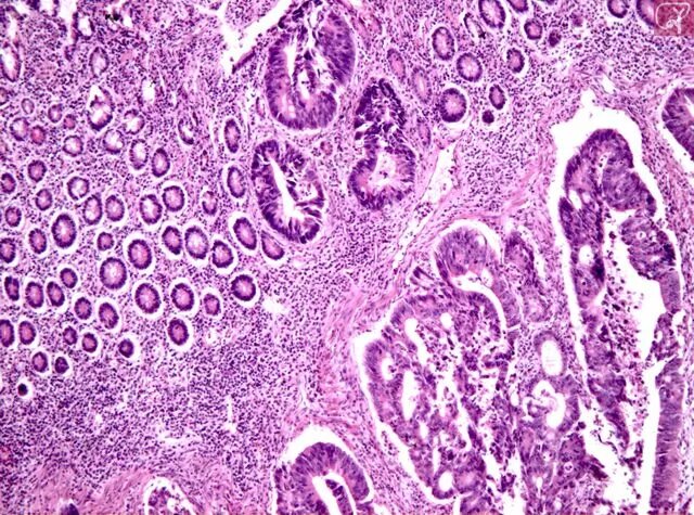
Хотя рак толстой кишки может передаваться по наследству, несколько эпидемиологических исследований показали, что риск рака толстой кишки во многом определяется факторами окружающей среды, такими как высокое потребление алкоголя, диета с низким содержанием клетчатки, диета с высоким содержанием жиров и курение табака [148,149].
Влияние АСТ на рак толстой кишки Было показано, что АСТ проявляет противораковое действие при различных типах рака, таких как рак желудка, рак молочной железы и рак простаты [94,153,154].
На нескольких моделях рака толстой кишки было показано, что АСТ регулирует такие признаки рака, как пролиферация, метастазирование и апоптоз [155].
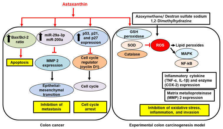
В клетках рака толстой кишки WiDr введение АСТ (1,25–250 мкМ; 72 ч) оказывало цитопротекторное действие путем ингибирования пролиферации клеток [156]. Аналогично, обработка богатым АСТ экстрактом Haematococcus pluvialis (25 мкл/мл; 24 часа) на клетках рака толстой кишки HCT-116 значительно увеличивала экспрессию p53, p21 и p27 и подавляла экспрессию циклина D1 и фосфорилирование AKT [157]. Кроме того, экстракт Haematococcus pluvialis увеличивал фосфорилирование p38, JNK и ERK1/2, одновременно изменяя соотношение Bax/Bcl-2. Эти результаты показывают, что АСТ может эффективно ингибировать рост клеток толстой кишки, ингибируя развитие клеточного цикла и усиливая апоптоз [157].
Лечение АСТ оказывается высокоэффективным при раке, связанном с колитом, который является относительно незначительным фактором риска. В мышиной модели канцерогенеза толстой кишки, опосредованной азоксиметаном (АОМ)/DSS, диетическое введение АСТ (200 частей на миллион; 17 недель) уменьшало пролиферативные поражения толстой кишки, возможно, за счет ингибирования экспрессии NF-κB, TNF-α и IL-1β. при злокачественных новообразованиях [43]. Танака и др. также сообщили о аналогичных результатах, где диета, содержащая АСТ (500 частей на миллион; 34 недели), заметно ингибировала развитие аберрантных очагов крипт посредством ингибирования активности пролиферации клеток [158]. У крыс, получавших 1,2-диметилгидразин (ДМГ) для индукции канцерогенеза толстой кишки, предварительная обработка АСТ (15 мг/кг массы тела; 16 недель) заметно уменьшала тяжесть поражений, а также количество аберрантных очагов крипт [159] . Механически у крыс, получавших АСТ, наблюдались повышенные уровни антиоксидантов, таких как СОД, КАТ и GPX, а также снижение уровня перекисного окисления липидов. В той же экспериментальной модели у крыс, которым вводили АСТ (15 мг/кг массы тела; 16 недель), наблюдалось значительное ингибирование экспрессии NF-κB-p65, ЦОГ-2, матриксной металлопротеиназы (ММП) 2/9, ядерного антигена пролиферирующих клеток ( PCNA) и ERK2, индуцированные DMH [160]. В соответствии с этими наблюдениями АСТ также уменьшал колоректальный канцерогенез, связанный с ожирением. Мышам C57BL/KsJ-db/db с нулевым рецептором лептина вводили АОМ, вызывающий спонтанное ожирение, Kochi et al. продемонстрировали, что диета, содержащая АСТ (200 ppm; восемь недель), снижает окислительный стресс, стимулируя экспрессию SOD1, GPX и CAT в слизистой оболочке толстой кишки [161]. У мышей db/db, получавших AST, также наблюдалось снижение количества клеток NF-κB+ и PCNA+ и снижение экспрессии генов IL-6, IL-1β, F4/80, хемокина (мотив CC), лиганда 2 (CCL2) и хемокина (C-X-C). мотив) лиганд 2 (CXCL2) в толстой кишке. Противораковая активность АСТ также может быть опосредована модуляцией микроРНК (миР). В клеточных линиях рака толстой кишки CT26 и HCT-116 обработка АСТ (50–100 мкМ; 24 часа) транскрипционно подавляла онкогенный транскрипционный фактор MYC и тем самым усиливала экспрессию миР-29а-3р и миР-200а, которые известны проявлять антиметастатический эффект [162]. В свою очередь, увеличение количества миР-29a-3p и миР-200а приводило к снижению экспрессии MMP2 и гомеобокса 1, связывающего E-бокс с цинковым пальцем (ZEB1), который опосредует эпителиально-мезенхимальный переход клеток колоректального рака. Эти механизмы были также подтверждены у голых мышей BALB/c, которым инъецировали клетки рака толстой кишки, где введение АСТ (25–50 мг/кг массы тела; четыре недели) ингибирует метастазирование рака толстой кишки в легкие через MYC/miR-29a-3p. и ось миР-200а [162]. В целом, эти данные свидетельствуют о том, что АСТ оказывает антиоксидантное, противовоспалительное и антиметастазное действие против прогрессирования рака толстой кишки.
АСТ может быть потенциальным кандидатом на роль химиопрофилактического средства заболевания.
Однако клинических испытаний на людях не проводилось.
На рис. 4 показан предполагаемый механизм ингибирующего действия АСТ на пролиферацию, метастазирование и окисление и стресс-опосредованное воспаление и инвазия в развитии и прогрессировании рака толстой кишки.
Кроме того, исследования четко установили молекулярные связи между хроническим воспалением, окислительным стрессом и колоректальным канцерогенезом [150,151]. Хотя молекулярные механизмы, с помощью которых АФК и воспаление способствуют канцерогенезу, все еще выясняются, редокс-чувствительные транскрипционные факторы, такие как NF-κB, AP-1 и STAT3, активно участвуют в канцерогенезе толстой кишки [152]. Таким образом, было идентифицировано, что различные химиопротекторные агенты, антиоксиданты и противовоспалительные биологически активные соединения обеспечивают молекулярную стратегию лечения рака толстой кишки путем модуляции аномально активированных NF-κB, AP-1 и STAT3.
6. Выводы
В этом обзоре антиоксидантные и противовоспалительные свойства АСТ были подчеркнуты на различных экспериментальных моделях язв и рака желудочно-кишечного тракта. Расположенный на поверхности и внутри клеточных мембран, АСТ действует как антиоксидант, надежно нейтрализуя АФК и липидные пероксильные радикалы [49,51]. Более того, АСТ также усиливает активность антиоксидантных ферментов, таких как СОД, КАТ и GPX [105,106,109]. В качестве модулятора воспаления АСТ регулирует воспалительные сигнальные пути, такие как NF-κB, AP-1 и МАРК, и одновременно подавляет экспрессию провоспалительных цитокинов, таких как IL-6, IL-1β, IL-8 и TNF-α. [43,92,144]. В моделях рака желудочно-кишечного тракта АСТ, по-видимому, ингибирует рост и метастазирование раковых клеток путем модуляции сигнальных путей, связанных с пролиферацией клеток, а также апоптоза, аутофагии и микроРНК [125,134,162]. Это было дополнительно подтверждено анализом RNA-Seq в клетках рака желудка, где профилирование выявило несколько генов-мишеней АСТ, которые связаны с пролиферацией и метастазированием раковых клеток [128,129]. Учитывая его безопасный профиль как биологически активного соединения [163], АСТ может стать эффективным и нетоксичным средством лечения этих заболеваний желудочно-кишечного тракта. Необходимо провести хорошо организованные клинические испытания для определения влияния АСТ на заболевания желудочно-кишечного тракта у людей.
References
1. Sies H., Belousov V.V., Chandel N.S., Davies M.J., Jones D.P., Mann G.E., Murphy M.P., Yamamoto M., Winterbourn C. Defining roles of specific reactive oxygen species (ROS) in cell biology and physiology. Nature Rev. Mol. Cell Biol. 2022;23:499–515. doi: 10.1038/s41580-022-00456-z. [PubMed] [CrossRef] [Google Scholar]
2. Bayr H. Reactive oxygen species. Critl. Care Med. 2005;33:S498–S501. doi: 10.1097/01.CCM.0000186787.64500.12. [PubMed] [CrossRef] [Google Scholar]
3. Pizzino G., Irrera N., Cucinotta M., Pallio G., Mannino F., Arcoraci V., Squadrito F., Altavilla D., Bitto A. Oxidative stress: Harms and benefits for human health. Oxid. Med. Cell. Longev. 2017;2017:8416763. doi: 10.1155/2017/8416763. [PMC free article] [PubMed] [CrossRef] [Google Scholar]
4. Brieger K., Schiavone S., Miller F.J., Krause K.-H. Reactive oxygen species: From health to disease. Swiss Med. Wkly. 2012;142:w13659. doi: 10.4414/smw.2012.13659. [PubMed] [CrossRef] [Google Scholar]
5. Ray P.D., Huang B.-W., Tsuji Y. Reactive oxygen species (ROS) homeostasis and redox regulation in cellular signaling. Cell Signal. 2012;24:981–990. doi: 10.1016/j.cellsig.2012.01.008. [PMC free article] [PubMed] [CrossRef] [Google Scholar]
6. Bondia-Pons I., Ryan L., Martinez J.A. Oxidative stress and inflammation interactions in human obesity. J. Physiol. Biochem. 2012;68:701–711. doi: 10.1007/s13105-012-0154-2. [PubMed] [CrossRef] [Google Scholar]
7. Biswas S.K. Does the interdependence between oxidative stress and inflammation explain the antioxidant paradox? Oxid. Med. Cell. Longev. 2016;2016:5698931. doi: 10.1155/2016/5698931. [PMC free article] [PubMed] [CrossRef] [Google Scholar]
8. Collins T. Robbins Pathologic Basis of Disease. Saunders; Philadelphia, PE, USA: 1999. Acute and chronic inflammation; pp. 83–84. [Google Scholar]
9. Essick E.E., Sam F. Oxidative stress and autophagy in cardiac disease, neurological disorders, aging and cancer. Oxid. Med. Cell. Longev. 2010;3:168–177. doi: 10.4161/oxim.3.3.12106. [PMC free article] [PubMed] [CrossRef] [Google Scholar]
10. Soybel D.I. Anatomy and physiology of the stomach. Surg. Clin. N. Am. 2005;85:875–894. doi: 10.1016/j.suc.2005.05.009. [PubMed] [CrossRef] [Google Scholar]
11. Suzuki H., Nishizawa T., Tsugawa H., Mogami S., Hibi T. Roles of oxidative stress in stomach disorders. J. Clin. Biochem. Nutr. 2012;50:35–39. doi: 10.3164/jcbn.11-115SR. [PMC free article] [PubMed] [CrossRef] [Google Scholar]
12. Gugliandolo E., Cordaro M., Fusco R., Peritore A.F., Siracusa R., Genovese T., D’Amico R., Impellizzeri D., Di Paola R., Cuzzocrea S. Protective effect of snail secretion filtrate against ethanol-induced gastric ulcer in mice. Sci. Rep. 2021;11:1–12. doi: 10.1038/s41598-021-83170-8. [PMC free article] [PubMed] [CrossRef] [Google Scholar]
13. Fox J.G., Wang T.C. Inflammation, atrophy, and gastric cancer. J. Clin. Investig. 2007;117:60–69. doi: 10.1172/JCI30111. [PMC free article] [PubMed] [CrossRef] [Google Scholar]
14. Correa P., Piazuelo M.B. The gastric precancerous cascade. J. Dig. Dis. 2012;13:2–9. doi: 10.1111/j.1751-2980.2011.00550.x. [PMC free article] [PubMed] [CrossRef] [Google Scholar]
15. Jena G., Trivedi P.P., Sandala B. Oxidative stress in ulcerative colitis: An old concept but a new concern. Free. Radic. Res. 2012;46:1339–1345. doi: 10.3109/10715762.2012.717692. [PubMed] [CrossRef] [Google Scholar]
16. Perše M. Oxidative stress in the pathogenesis of colorectal cancer: Cause or consequence? BioMed Res. Int. 2013;2013:725710. doi: 10.1155/2013/725710. [PMC free article] [PubMed] [CrossRef] [Google Scholar]
17. Sørbye H., Svanes K. The role of blood flow in gastric mucosal defence, damage and healing. Dig. Dis. 1994;12:305–317. doi: 10.1159/000171465. [PubMed] [CrossRef] [Google Scholar]
18. Warzecha Z., Dembiński A., Brzozowski T., Ceranowicz P., Pajdo R., Niemiec J., Drozdowicz D., Mitis-Musioł M., Konturek S.J. Gastroprotective effect of histamine and acid secretion on ammonia-induced gastric lesions in rats. Scand J. Gastroenterol. 2000;35:916–924. doi: 10.1080/003655200750022959. [PubMed] [CrossRef] [Google Scholar]
19. Warzecha Z., Dembiński A., Brzozowski T., Ceranowicz P., Dembiński M., Stachura J., Konturek S.J. Histamine in stress ulcer prophylaxis in rats. J. Physiol. Pharmacol. 2001;52:407–421. [PubMed] [Google Scholar]
20. Konarska K., Cieszkowski J., Warzecha Z., Ceranowicz P., Chmura A., Kuśnierz-Cabala B., Gałązka K., Kowalczyk P.I., Miskiewicz A., Konturek T.J., et al. Treatment with obestatin-a ghrelin gene-encoded peptide-reduces the severity of experimental colitis evoked by trinitrobenzene sulfonic acid. Int. J. Mol. Sci. 2018;19:1643. doi: 10.3390/ijms19061643. [PMC free article] [PubMed] [CrossRef] [Google Scholar]
21. Dembiński A., Warzecha Z., Ceranowicz P., Dembiński M., Cieszkowski J., Gosiewski T., Bulanda M., Kuśnierz-Cabala B., Gałązka K., Konturek P.C. Synergic interaction of Rifaximin and Mutaflor (Escherichia coli Nissle 1917) in the treatment of acetic acid-induced colitis in rats. Gastroenterol. Res. Pract. 2016;2016:3126280. doi: 10.1155/2016/3126280. [PMC free article] [PubMed] [CrossRef] [Google Scholar]
22. Granger D.N., Kvietys P.R. Reperfusion injury and reactive oxygen species: The evolution of a concept. Redox Biol. 2015;6:524–551. doi: 10.1016/j.redox.2015.08.020. [PMC free article] [PubMed] [CrossRef] [Google Scholar]
23. Li Y., Feng D., Wang Z., Zhao Y., Sun R., Tian D., Liu D., Zhang F., Ning S., Yao J., et al. Ischemia-induced ACSL4 activation contributes to ferroptosis-mediated tissue injury in intestinal ischemia/reperfusion. Cell Death Differ. 2019;26:2284–2299. doi: 10.1038/s41418-019-0299-4. [PMC free article] [PubMed] [CrossRef] [Google Scholar]
24. Akki R., Raghay K., Errami M. Potentiality of ghrelin as antioxidant and protective agent. Redox Rep. 2021;26:71–79. doi: 10.1080/13510002.2021.1913374. [PMC free article] [PubMed] [CrossRef] [Google Scholar]
25. Ginter G., Ceranowicz P., Warzecha Z. Protective and healing effects of ghrelin and risk of cancer in the digestive system. Int. J. Mol. Sci. 2021;22:10571. doi: 10.3390/ijms221910571. [PMC free article] [PubMed] [CrossRef] [Google Scholar]
26. Warzecha Z., Ceranowicz P., Dembinski M., Cieszkowski J., Ginter G., Ptak-Belowska A., Dembinski A. Involvement of cyclooxygenase-1 and cyclooxygenase-2 activity in the therapeutic effect of ghrelin in the course of ethanol-induced gastric ulcers in rats. J. Physiol. Pharmacol. 2014;65:95–106. [PubMed] [Google Scholar]
27. Sibilia V., Rindi G., Pagani F., Rapetti D., Locatelli V., Torsello A., Campanini N., Deghenghi R., Netti C. Ghrelin protects against ethanol-induced gastric ulcers in rats: Studies on the mechanisms of action. Endocrinology. 2003;144:353–359. doi: 10.1210/en.2002-220756. [PubMed] [CrossRef] [Google Scholar]
28. Ceranowicz P., Warzecha Z., Dembinski A., Sendur R., Cieszkowski J., Ceranowicz D., Pawlik W.W., Kuwahara A., Kato I., Konturek P.C. Treatment with ghrelin accelerates the healing of acetic acid-induced gastric and duodenal ulcers in rats. J. Physiol. Pharmacol. 2009;60:87–98. [PubMed] [Google Scholar]
29. Stempniewicz A., Ceranowicz P., Warzecha Z. Potential therapeutic effects of gut hormones, ghrelin and obestatin in oral mucositis. Int. J. Mol. Sci. 2019;20:1534. doi: 10.3390/ijms20071534. [PMC free article] [PubMed] [CrossRef] [Google Scholar]
30. Dembinski A., Warzecha Z., Ceranowicz P., Tomaszewska R., Stachura J., Konturek S.J., Konturek P.C. Ghrelin attenuates the development of acute pancreatitis in rat. J. Physiol. Pharmacol. 2003;54:561–573. [PubMed] [Google Scholar]
31. Dembinski A., Warzecha Z., Ceranowicz P., Cieszkowski J., Pawlik W.W., Tomaszewska P., Kuśnierz-Cabala B., Naskalski J.W., Kuwahara A., Kato I. Role of growth hormone and insulin-like growth factor-1 in the protective effect of ghrelin in ischemia/reperfusion-induced acute pancreatitis. Growth Horm. IGF Res. 2006;16:348–356. doi: 10.1016/j.ghir.2006.09.003. [PubMed] [CrossRef] [Google Scholar]
32. Maduzia D., Matuszyk A., Ceranowicz D., Warzecha Z., Ceranowicz P., Fyderek K., Galazka K., Dembinski A. The influence of pretreatment with ghrelin on the development of acetic-acid-induced colitis in rats. J. Physiol. Pharmacol. 2015;66:875–885. [PubMed] [Google Scholar]
33. Matuszyk A., Ceranowicz P., Warzecha Z., Cieszkowski J., Ceranowicz D., Gałązka K., Bonior J., Jaworek J., Bartuś K., Gil K., et al. Exogenous ghrelin accelerates the healing of acetic acid-induced colitis in rats. Int. J. Mol. Sci. 2016;17:1455. doi: 10.3390/ijms17091455. [PMC free article] [PubMed] [CrossRef] [Google Scholar]
34. 34 Bukowczan J., Warzecha Z., Ceranowicz P., Kusnierz-Cabala B., Tomaszewska R., Dembinski A. Therapeutic effect of ghrelin in the course of ischemia/reperfusion-induced acute pancreatitis. Curr. Pharm. Des. 2015;21:2284–2290. doi: 10.2174/1381612821666150105152553. [PubMed] [CrossRef] [Google Scholar]
35. Warzecha Z., Ceranowicz D., Dembiński A., Ceranowicz P., Cieszkowski J., Kuwahara A., Kato I., Dembiński M., Konturek P.C. Ghrelin accelerates the healing of cysteamine-induced duodenal ulcers in rats. Med. Sci. Monit. 2012;18:BR181–BR187. doi: 10.12659/MSM.882727. [PMC free article] [PubMed] [CrossRef] [Google Scholar]
36. Matuszyk A., Ceranowicz D., Warzecha Z., Ceranowicz P., Fyderek K., Gałązka K., Cieszkowski J., Bonior J., Jaworek J., Pihut M., et al. The influence of ghrelin on the development of dextran sodium sulfate-induced colitis in rats. BioMed Res. Int. 2015;2015:718314. doi: 10.1155/2015/718314. [PMC free article] [PubMed] [CrossRef] [Google Scholar]
37. Ceranowicz P., Warzecha Z., Cieszkowski J., Ceranowicz D., Kuśnierz-Cabala B., Bonior J., Jaworek J., Ambroży T., Gil K., Olszanecki R., et al. Essential role of growth hormone and IGF-1 in therapeutic effect of ghrelin in the course of acetic acid-induced colitis. Int. J. Mol. Sci. 2017;18:1118. doi: 10.3390/ijms18061118. [PMC free article] [PubMed] [CrossRef] [Google Scholar]
38. Matuszyk A., Ceranowicz P., Warzecha Z., Cieszkowski J., Gałązka K., Bonior J., Jaworek J., Konturek P.C., Gil K., Dembinski A. Pretreatment with obestatin inhibits the development of acetic acid-induced colitis in rats. Arch. Med. Sci. 2018;14:920–929. doi: 10.5114/aoms.2016.58749. [PMC free article] [PubMed] [CrossRef] [Google Scholar]
39. Al-Bulishi M.S.M., Changhu X., Tang Q.-J. Health aspects of astaxanthin: A review. Canad. J. Clin. Nutr. 2015;3:71–78. doi: 10.14206/canad.j.clin.nutr.2015.02.08. [CrossRef] [Google Scholar]
40. Ushakumari U.N., Ramanujan R. Isolation of astaxanthin from marine yeast and study of its pharmacological activity. Int. Curr. Pharm. J. 2013;2:67–69. doi: 10.3329/icpj.v2i3.13584. [CrossRef] [Google Scholar]
41. Guerin M., Huntley M.E., Olaizola M. Haematococcus astaxanthin: Applications for human health and nutrition. Trends Biotechnol. 2003;21:210–216. doi: 10.1016/S0167-7799(03)00078-7. [PubMed] [CrossRef] [Google Scholar]
42. Bennedsen M., Wang X., Willén R., Wadström T., Andersen L.P. Treatment of H. pylori infected mice with antioxidant astaxanthin reduces gastric inflammation, bacterial load and modulates cytokine release by splenocytes. Immunol. Lett. 2000;70:185–189. doi: 10.1016/S0165-2478(99)00145-5. [PubMed] [CrossRef] [Google Scholar]
43. Yasui Y., Hosokawa M., Mikami N., Miyashita K., Tanaka T. Dietary astaxanthin inhibits colitis and colitis-associated colon carcinogenesis in mice via modulation of the inflammatory cytokines. Chem. Biol. Interact. 2011;193:79–87. doi: 10.1016/j.cbi.2011.05.006. [PubMed] [CrossRef] [Google Scholar]
44. Naguib Y.M. Antioxidant activities of astaxanthin and related carotenoids. J. Agric. Food Chem. 2000;48:1150–1154. doi: 10.1021/jf991106k. [PubMed] [CrossRef] [Google Scholar]
45. Rao A.R., Baskaran V., Sarada R., Ravishankar G.A. In vivo bioavailability and antioxidant activity of carotenoids from microalgal biomass—A repeated dose study. Food Res. Int. 2013;54:711–717. [Google Scholar]
46. Stahl W., Sies H. Antioxidant activity of carotenoids. Mol. Asp. Med. 2003;24:345–351. doi: 10.1016/S0098-2997(03)00030-X. [PubMed] [CrossRef] [Google Scholar]
47. McNulty H., Jacob R.F., Mason R.P. Biologic activity of carotenoids related to distinct membrane physicochemical interactions. Am. J. Cardiol. 2008;101:S20–S29. doi: 10.1016/j.amjcard.2008.02.004. [PubMed] [CrossRef] [Google Scholar]
48. Goto S., Kogure K., Abe K., Kimata Y., Kitahama K., Yamashita E., Terada H. Efficient radical trapping at the surface and inside the phospholipid membrane is responsible for highly potent antiperoxidative activity of the carotenoid astaxanthin. Bioch. Biophys. Acta (BBA)-Biomembr. 2001;1512:251–258. doi: 10.1016/S0005-2736(01)00326-1. [PubMed] [CrossRef] [Google Scholar]
49. Pereira C.P.M., Souza A.C.R., Vasconcelos A.R., Prado P.S. Antioxidant and anti-inflammatory mechanisms of action of astaxanthin in cardiovascular diseases. Int. J. Mol. Med. 2021;47:37–48. doi: 10.3892/ijmm.2020.4783. [PMC free article] [PubMed] [CrossRef] [Google Scholar]
50. Hussein G., Sankawa U., Goto H., Matsumoto K., Watanabe H. Astaxanthin, a carotenoid with potential in human health and nutrition. J. Nat. Prod. 2006;69:443–449. doi: 10.1021/np050354+. [PubMed] [CrossRef] [Google Scholar]
51. Barros M.P., Pinto E., Colepicolo P., Pedersén M. Astaxanthin and peridinin inhibit oxidative damage in Fe2+-loaded liposomes: Scavenging oxyradicals or changing membrane permeability? Biochem. Biophys. Res. Commun. 2001;288:225–232. doi: 10.1006/bbrc.2001.5765. [PubMed] [CrossRef] [Google Scholar]
52. McNulty H.P., Byun J., Lockwood S.F., Jacob R.F., Mason R.P. Differential effects of carotenoids on lipid peroxidation due to membrane interactions: X-ray diffraction analysis. Bioch. Biophys. Acta (BBA)-Biomembr. 2007;1768:167–174. doi: 10.1016/j.bbamem.2006.09.010. [PubMed] [CrossRef] [Google Scholar]
53. Pashkow F.J., Watumull D.G., Campbell C.L. Astaxanthin: A novel potential treatment for oxidative stress and inflammation in cardiovascular disease. Am. J. Cardiol. 2008;101:S58–S68. doi: 10.1016/j.amjcard.2008.02.010. [PubMed] [CrossRef] [Google Scholar]
54. May J.M. Is ascorbic acid an antioxidant for the plasma membrane? FASEB J. 1999;13:995–1006. doi: 10.1096/fasebj.13.9.995. [PubMed] [CrossRef] [Google Scholar]
55. Dose J., Matsugo S., Yokokawa H., Koshida Y., Okazaki S., Seidel U., Eggersdorfer M., Rimbach G., Esatbeyoglu T. Free radical scavenging and cellular antioxidant properties of astaxanthin. Int. J. Mol. Sci. 2016;17:103. doi: 10.3390/ijms17010103. [PMC free article] [PubMed] [CrossRef] [Google Scholar]
56. Kohandel Z., Farkhondeh T., Aschner M., Pourbagher-Shahri A.M., Samarghandian S. Anti-inflammatory action of astaxanthin and its use in the treatment of various diseases. Biomed. Pharmacother. 2022;145:112179. doi: 10.1016/j.biopha.2021.112179. [PubMed] [CrossRef] [Google Scholar]
57. Speranza L., Pesce M., Patruno A., Franceschelli S., De Lutiis M.A., Grilli A., Felaco M. Astaxanthin treatment reduced oxidative induced pro-inflammatory cytokines secretion in U937: SHP-1 as a novel biological target. Mar. Drugs. 2012;10:890–899. doi: 10.3390/md10040890. [PMC free article] [PubMed] [CrossRef] [Google Scholar]
58. Suzuki Y., Ohgami K., Shiratori K., Jin X.-H., Ilieva I., Koyama Y., Yazawa K., Yoshida K., Kase S., Ohno S. Suppressive effects of astaxanthin against rat endotoxin-induced uveitis by inhibiting the NF-κB signaling pathway. Exp. Eye Res. 2006;82:275–281. doi: 10.1016/j.exer.2005.06.023. [PubMed] [CrossRef] [Google Scholar]
59. Li J., Wang F., Xia Y., Dai W., Chen K., Li S., Liu T., Zheng Y., Wang J., Lu W. Astaxanthin pretreatment attenuates hepatic ischemia reperfusion-induced apoptosis and autophagy via the ROS/MAPK pathway in mice. Mar. Drugs. 2015;13:3368–3387. doi: 10.3390/md13063368. [PMC free article] [PubMed] [CrossRef] [Google Scholar]
60. Yang X., Guo A.-L., Pang Y.-P., Cheng X.-J., Xu T., Li X.-R., Liu J., Zhang Y.-Y., Liu Y. Astaxanthin attenuates environmental tobacco smoke-induced cognitive deficits: A critical role of p38 MAPK. Mar. Drugs. 2019;17:24. doi: 10.3390/md17010024. [PMC free article] [PubMed] [CrossRef] [Google Scholar]
61. Liu G., Shi Y., Peng X., Liu H., Peng Y., He L. Astaxanthin attenuates adriamycin-induced focal segmental glomerulosclerosis. Pharmacology. 2015;95:193–200. doi: 10.1159/000381314. [PubMed] [CrossRef] [Google Scholar]
62. Xu L., Zhu J., Yin W., Ding X. Astaxanthin improves cognitive deficits from oxidative stress, nitric oxide synthase and inflammation through upregulation of PI3K/Akt in diabetes rat. Int. J. Clin. Exp. Pathol. 2015;8:6083. [PMC free article] [PubMed] [Google Scholar]
63. Yuan J.P., Peng J., Yin K., Wang J.H. Potential health-promoting effects of astaxanthin: A high-value carotenoid mostly from microalgae. Mol. Nutr. Food Res. 2011;55:150–165. doi: 10.1002/mnfr.201000414. [PubMed] [CrossRef] [Google Scholar]
64. Ambati R.R., Siew Moi P., Ravi S., Aswathanarayana R.G. Astaxanthin: Sources, extraction, stability, biological activities and its commercial applications—A review. Mar. Drugs. 2014;12:128–152. doi: 10.3390/md12010128. [PMC free article] [PubMed] [CrossRef] [Google Scholar]
65. Sy C., Gleize B., Dangles O., Landrier J.F., Veyrat C.C., Borel P. Effects of physicochemical properties of carotenoids on their bioaccessibility, intestinal cell uptake, and blood and tissue concentrations. Mol. Nutr. Food Res. 2012;56:1385–1397. doi: 10.1002/mnfr.201200041. [PubMed] [CrossRef] [Google Scholar]
66. Reboul E., Abou L., Mikail C., Ghiringhelli O., André M., Portugal H., Jourdheuil-Rahmani D., Amiot M.-J., Lairon D., Borel P. Lutein transport by Caco-2 TC-7 cells occurs partly by a facilitated process involving the scavenger receptor class B type I (SR-BI) Biochem. J. 2005;387:455–461. doi: 10.1042/BJ20040554. [PMC free article] [PubMed] [CrossRef] [Google Scholar]
67. Kiefer C., Sumser E., Wernet M.F., Von Lintig J. A class B scavenger receptor mediates the cellular uptake of carotenoids in Drosophila. Proc. Nat. Acad. Sci. USA. 2002;99:10581–10586. doi: 10.1073/pnas.162182899. [PMC free article] [PubMed] [CrossRef] [Google Scholar]
68. During A., Dawson H.D., Harrison E.H. Carotenoid transport is decreased and expression of the lipid transporters SR-BI, NPC1L1, and ABCA1 is downregulated in Caco-2 cells treated with ezetimibe. J. Nutr. 2005;135:2305–2312. doi: 10.1093/jn/135.10.2305. [PubMed] [CrossRef] [Google Scholar]
69. Zhou Q., Xu J., Yang L., Gu C., Xue C. Thermal stability and oral absorbability of astaxanthin esters from Haematococcus pluvialis in Balb/c mice. J. Sci. Food Agric. 2019;99:3662–3671. doi: 10.1002/jsfa.9588. [PubMed] [CrossRef] [Google Scholar]
70. Petri D., Lundebye A.-K. Tissue distribution of astaxanthin in rats following exposure to graded levels in the feed. Comp. Biochem. Physiol. C Toxicol. Pharmacol. 2007;145:202–209. doi: 10.1016/j.cbpc.2006.12.008. [PubMed] [CrossRef] [Google Scholar]
71. Singh G.S., Ismail M., Zulkefli N., Affandi M., Meor M. Tissue distribution of astaxanthin formulation in rats. Curr. Nutr. Food Sci. 2018;14:329–334. doi: 10.2174/1573401313666170614092146. [CrossRef] [Google Scholar]
72. Østerlie M., Bjerkeng B., Liaaen-Jensen S. Plasma appearance and distribution of astaxanthin E/Z and R/S isomers in plasma lipoproteins of men after single dose administration of astaxanthin. J. Nutr. Biochem. 2000;11:482–490. doi: 10.1016/S0955-2863(00)00104-2. [PubMed] [CrossRef] [Google Scholar]
73. Coral-Hinostroza G.N., Ytrestøyl T., Ruyter B., Bjerkeng B. Plasma appearance of unesterified astaxanthin geometrical E/Z and optical R/S isomers in men given single doses of a mixture of optical 3 and 3′ R/S isomers of astaxanthin fatty acyl diesters. Comp. Biochem. Physiol. C Toxicol. Pharmacol. 2004;139:99–110. doi: 10.1016/j.cca.2004.09.011. [PubMed] [CrossRef] [Google Scholar]
74. Malfertheiner P., Chan F.K., McColl K.E. Peptic ulcer disease. Lancet. 2009;374:1449–1461. doi: 10.1016/S0140-6736(09)60938-7. [PubMed] [CrossRef] [Google Scholar]
75. Lanas A., Chan F.K. Peptic ulcer disease. Lancet. 2017;390:613–624. doi: 10.1016/S0140-6736(16)32404-7. [PubMed] [CrossRef] [Google Scholar]
76. Mittal M., Siddiqui M.R., Tran K., Reddy S.P., Malik A.B. Reactive oxygen species in inflammation and tissue injury. Antioxid. Redox Signal. 2014;20:1126–1167. doi: 10.1089/ars.2012.5149. [PMC free article] [PubMed] [CrossRef] [Google Scholar]
77. Bhattacharyya A., Chattopadhyay R., Mitra S., Crowe S.E. Oxidative stress: An essential factor in the pathogenesis of gastrointestinal mucosal diseases. Physiol. Rev. 2014;94:329–354. doi: 10.1152/physrev.00040.2012. [PMC free article] [PubMed] [CrossRef] [Google Scholar]
78. Panday A., Sahoo M.K., Osorio D., Batra S. NADPH oxidases: An overview from structure to innate immunity-associated pathologies. Cell Mol. Immunol. 2015;12:5–23. doi: 10.1038/cmi.2014.89. [PMC free article] [PubMed] [CrossRef] [Google Scholar]
79. Fujioka S., Niu J., Schmidt C., Sclabas G.M., Peng B., Uwagawa T., Li Z., Evans D.B., Abbruzzese J.L., Chiao P.J. NF-κB and AP-1 connection: Mechanism of NF-κB-dependent regulation of AP-1 activity. Mol. Cell. Biol. 2004;24:7806–7819. doi: 10.1128/MCB.24.17.7806-7819.2004. [PMC free article] [PubMed] [CrossRef] [Google Scholar]
80. Handa O., Naito Y., Yoshikawa T. Helicobacter pylori: A ROS-inducing bacterial species in the stomach. Inflamm. Res. 2010;59:997–1003. doi: 10.1007/s00011-010-0245-x. [PubMed] [CrossRef] [Google Scholar]
81. Iwamoto T., Hosoda K., Hirano R., Kurata H., Matsumoto A., Miki W., Kamiyama M., Itakura H., Yamamoto S., Kondo K. Inhibition of low-density lipoprotein oxidation by astaxanthin. J. Atheroscler. Thromb. 2000;7:216–222. doi: 10.5551/jat1994.7.216. [PubMed] [CrossRef] [Google Scholar]
82. Davies G., Simmonds N., Stevens T., Sheaff M., Banatvala N., Laurenson I., Blake D., Rampton D. Helicobacter pylori stimulates antral mucosal reactive oxygen metabolite production in vivo. Gut. 1994;35:179–185. doi: 10.1136/gut.35.2.179. [PMC free article] [PubMed] [CrossRef] [Google Scholar]
83. Lee K.E., Khoi P.N., Xia Y., Park J.S., Joo Y.E., Kim K.K., Choi S.Y., Do Jung Y. Helicobacter pylori and interleukin-8 in gastric cancer. World J. Gastroenterol. 2013;19:8192. doi: 10.3748/wjg.v19.i45.8192. [PMC free article] [PubMed] [CrossRef] [Google Scholar]
84. Jang S., Jones K.R., Olsen C.H., Joo Y.M., Yoo Y.-J., Chung I.-S., Cha J.-H., Merrell D.S. Epidemiological link between gastric disease and polymorphisms in VacA and CagA. J. Clin. Microbiol. 2010;48:559–567. doi: 10.1128/JCM.01501-09. [PMC free article] [PubMed] [CrossRef] [Google Scholar]
85. Censini S., Lange C., Xiang Z., Crabtree J.E., Ghiara P., Borodovsky M., Rappuoli R., Covacci A. cag, a pathogenicity island of Helicobacter pylori, encodes type I-specific and disease-associated virulence factors. Proc. Nat. Acad. Sci. USA. 1996;93:14648–14653. doi: 10.1073/pnas.93.25.14648. [PMC free article] [PubMed] [CrossRef] [Google Scholar]
86. Aihara M., Tsuchimoto D., Takizawa H., Azuma A., Wakebe H., Ohmoto Y., Imagawa K., Kikuchi M., Mukaida N., Matsushima K. Mechanisms involved in Helicobacter pylori-induced interleukin-8 production by a gastric cancer cell line, MKN45. Infect Immun. 1997;65:3218–3224. doi: 10.1128/iai.65.8.3218-3224.1997. [PMC free article] [PubMed] [CrossRef] [Google Scholar]
87. Yamaoka Y., Kikuchi S., El–Zimaity H.M., Gutierrez O., Osato M.S., Graham D.Y. Importance of Helicobacter pylori oipA in clinical presentation, gastric inflammation, and mucosal interleukin 8 production. Gastroenterology. 2002;123:414–424. doi: 10.1053/gast.2002.34781. [PubMed] [CrossRef] [Google Scholar]
88. Crabtree J., Covacci A., Farmery S., Xiang Z., Tompkins D., Perry S., Lindley I., Rappuoli R. Helicobacter pylori induced interleukin-8 expression in gastric epithelial cells is associated with CagA positive phenotype. J. Clin. Pathol. 1995;48:41–45. doi: 10.1136/jcp.48.1.41. [PMC free article] [PubMed] [CrossRef] [Google Scholar]
89. Epplein M., Xiang Y.-B., Cai Q., Peek R.M., Li H., Correa P., Gao J., Wu J., Michel A., Pawlita M. Circulating cytokines and gastric cancer risk. Cancer Causes Control. 2013;24:2245–2250. doi: 10.1007/s10552-013-0284-z. [PMC free article] [PubMed] [CrossRef] [Google Scholar]
90. Ma J., Wu D., Hu X., Li J., Cao M., Dong W. Associations between cytokine gene polymorphisms and susceptibility to Helicobacter pylori infection and Helicobacter pylori related gastric cancer, peptic ulcer disease: A meta-analysis. PLoS ONE. 2017;12:e0176463. doi: 10.1371/journal.pone.0176463. [PMC free article] [PubMed] [CrossRef] [Google Scholar]
91. Atherton J.C., Blaser M.J. Coadaptation of Helicobacter pylori and humans: Ancient history, modern implications. J. Clin. Investig. 2009;119:2475–2487. doi: 10.1172/JCI38605. [PMC free article] [PubMed] [CrossRef] [Google Scholar]
92. Kim S.H., Lim J.W., Kim H. Astaxanthin inhibits mitochondrial dysfunction and interleukin-8 expression in Helicobacter pylori-infected gastric epithelial cells. Nutrients. 2018;10:1320. doi: 10.3390/nu10091320. [PMC free article] [PubMed] [CrossRef] [Google Scholar]
93. Kim S.H., Lim J.W., Kim H. Astaxanthin prevents decreases in superoxide dismutase 2 level and superoxide dismutase activity in helicobacter pylori-infected gastric epithelial cells. J. Cancer Preven. 2019;24:54. doi: 10.15430/JCP.2019.24.1.54. [PMC free article] [PubMed] [CrossRef] [Google Scholar]
94. Han H., Lim J.W., Kim H. Astaxanthin inhibits Helicobacter pylori-induced inflammatory and oncogenic responses in gastric mucosal tissues of mice. J. Cancer Preven. 2020;25:244. doi: 10.15430/JCP.2020.25.4.244. [PMC free article] [PubMed] [CrossRef] [Google Scholar]
95. Chang W.-J., Du Y., Zhao X., Ma L.-Y., Cao G.-W. Inflammation-related factors predicting prognosis of gastric cancer. World J. Gastroenterol. 2014;20:4586. doi: 10.3748/wjg.v20.i16.4586. [PMC free article] [PubMed] [CrossRef] [Google Scholar]
96. Knutson K.L., Disis M. Tumor antigen-specific T helper cells in cancer immunity and immunotherapy. Cancer Immunol. Immunother. 2005;54:721–728. doi: 10.1007/s00262-004-0653-2. [PubMed] [CrossRef] [Google Scholar]
97. Whary M., Morgan T., Dangler C., Gaudes K., Taylor N., Fox J. Chronic active hepatitis induced by Helicobacter hepaticus in the A/JCr mouse is associated with a Th1 cell-mediated immune response. Infect Immun. 1998;66:3142–3148. doi: 10.1128/IAI.66.7.3142-3148.1998. [PMC free article] [PubMed] [CrossRef] [Google Scholar]
98. Davinelli S., Melvang H.M., Andersen L.P., Scapagnini G., Nielsen M.E. Astaxanthin from shrimp cephalothorax stimulates the immune response by enhancing IFN-γ, IL-10, and IL-2 secretion in splenocytes of Helicobacter pylori-infected mice. Mar. Drugs. 2019;17:382. doi: 10.3390/md17070382. [PMC free article] [PubMed] [CrossRef] [Google Scholar]
99. Andersen L.P., Holck S., Kupcinskas L., Kiudelis G., Jonaitis L., Janciauskas D., Permin H., Wadström T. Gastric inflammatory markers and interleukins in patients with functional dyspepsia treated with astaxanthin. FEMS Immunol. Med. Microbiol. 2007;50:244–248. doi: 10.1111/j.1574-695X.2007.00257.x. [PubMed] [CrossRef] [Google Scholar]
100. Park J.S., Chyun J.H., Kim Y.K., Line L.L., Chew B.P. Astaxanthin decreased oxidative stress and inflammation and enhanced immune response in humans. Nutr. Metabol. 2010;7:1–10. doi: 10.1186/1743-7075-7-18. [PMC free article] [PubMed] [CrossRef] [Google Scholar]
101. Cashman J.N. The mechanisms of action of NSAIDs in analgesia. Drugs. 1996;52:13–23. doi: 10.2165/00003495-199600525-00004. [PubMed] [CrossRef] [Google Scholar]
102. Tenenbaum J. The epidemiology of nonsteroidal anti-inflammatory drugs. Canad. J. Gastroenterol. 1999;13:119–122. doi: 10.1155/1999/361651. [PubMed] [CrossRef] [Google Scholar]
103. Calhoun W., Gilman S., Datko L., Copenhaver T., Carlson R. Interaction studies of tilomisole, aspirin, and naproxen in acute and chronic inflammation with assessment of gastrointestinal irritancy in the rat. Agents Actions. 1992;36:99–106. doi: 10.1007/BF01991236. [PubMed] [CrossRef] [Google Scholar]
104. Graham D.Y., Malaty H.M. Alendronate and naproxen are synergistic for development of gastric ulcers. Arch Intern. Med. 2001;161:107–110. doi: 10.1001/archinte.161.1.107. [PubMed] [CrossRef] [Google Scholar]
105. Kim J.-H., Kim Y.-S., Song G.-G., Park J.-J., Chang H.-I. Protective effect of astaxanthin on naproxen-induced gastric antral ulceration in rats. Eur. J. Pharmacol. 2005;514:53–59. doi: 10.1016/j.ejphar.2005.03.034. [PubMed] [CrossRef] [Google Scholar]
106. Kim J.-H., Choi S.-K., Lim W.-J., Chang H.-I. Protective effect of astaxanthin produced by Xanthophyllomyces dendrorhous mutant on indomethacin-induced gastric mucosal injury in rats. J. Microbiol. Biotechnol. 2004;14:996–1003. [Google Scholar]
107. Laksitorini M.D., Yathindranath V., Xiong W., Parkinson F.E., Thliveris J.A., Miller D.W. Impact of Wnt/β-catenin signaling on ethanol-induced changes in brain endothelial cell permeability. J. Neurochem. 2021;157:1118–1137. doi: 10.1111/jnc.15203. [PubMed] [CrossRef] [Google Scholar]
108. Brătucu M.N., Prunoiu V.-M., Strâmbu V., Brătucu E., Răvaş M.-M., Simion L., Petre R. Unusual Complicated Gastric Ulcers. Medicina. 2021;57:1345. doi: 10.3390/medicina57121345. [PMC free article] [PubMed] [CrossRef] [Google Scholar]
109. Kim J.-H., Choi S.-K., Choi S.-Y., Kim H.-K., Chang H.-I. Suppressive effect of astaxanthin isolated from the Xanthophyllomyces dendrorhous mutant on ethanol-induced gastric mucosal injury in rats. Biosci. Biotechnol. Biochem. 2005;69:1300–1305. doi: 10.1271/bbb.69.1300. [PubMed] [CrossRef] [Google Scholar]
110. Kamath B.S., Srikanta B.M., Dharmesh S.M., Sarada R., Ravishankar G.A. Ulcer preventive and antioxidative properties of astaxanthin from Haematococcus pluvialis. Eur. J. Pharmacol. 2008;590:387–395. doi: 10.1016/j.ejphar.2008.06.042. [PubMed] [CrossRef] [Google Scholar]
111. Murata K., Oyagi A., Takahira D., Tsuruma K., Shimazawa M., Ishibashi T., Hara H. Protective effects of astaxanthin from Paracoccus carotinifaciens on murine gastric ulcer models. Phytother. Res. 2012;26:1126–1132. doi: 10.1002/ptr.3681. [PubMed] [CrossRef] [Google Scholar]
112. Nishikawa Y., Minenaka Y., Ichimura M., Tatsumi K., Nadamoto T., Urabe K. Effects of astaxanthin and vitamin C on the prevention of gastric ulcerations in stressed rats. J. Nutr. Sci. Vitaminol. 2005;51:135–141. doi: 10.3177/jnsv.51.135. [PubMed] [CrossRef] [Google Scholar]
113. Van Cutsem E., Sagaert X., Topal B., Haustermans K., Prenen H. Gastric cancer. Lancet. 2016;388:2654–2664. doi: 10.1016/S0140-6736(16)30354-3. [PubMed] [CrossRef] [Google Scholar]
114. Ferlay J., Soerjomataram I., Dikshit R., Eser S., Mathers C., Rebelo M., Parkin D.M., Forman D., Bray F. Cancer incidence and mortality worldwide: Sources, methods and major patterns in GLOBOCAN 2012. Int. J. Cancer. 2015;136:E359–E386. doi: 10.1002/ijc.29210. [PubMed] [CrossRef] [Google Scholar]
115. Lian S., Li S., Zhu J., Xia Y., Do Jung Y. Nicotine stimulates IL-8 expression via ROS/NF-κB and ROS/MAPK/AP-1 axis in human gastric cancer cells. Toxicology. 2022;466:153062. doi: 10.1016/j.tox.2021.153062. [PubMed] [CrossRef] [Google Scholar]
116. Kathuria S., Mahadevan N., Balakumar P. Possible involvement of PPARγ-associated eNOS signaling activation in rosuvastatin-mediated prevention of nicotine-induced experimental vascular endothelial abnormalities. Mol. Cell. Biochem. 2013;374:61–72. doi: 10.1007/s11010-012-1505-6. [PubMed] [CrossRef] [Google Scholar]
117. Hamraz M., Salimi M., Esfahani M., Sedaghati B., Aslani H.R., Bazarchi A. Nicotine effect on P-ERK, COX-2, PGE2 and VEGF expression in oral squamous cancer. J. Biotechnol. 2010;150:442. doi: 10.1016/j.jbiotec.2010.09.631. [CrossRef] [Google Scholar]
118. Dasgupta P., Rizwani W., Pillai S., Kinkade R., Kovacs M., Rastogi S., Banerjee S., Carless M., Kim E., Coppola D. Nicotine induces cell proliferation, invasion and epithelial-mesenchymal transition in a variety of human cancer cell lines. Int. J. Cancer. 2009;124:36–45. doi: 10.1002/ijc.23894. [PMC free article] [PubMed] [CrossRef] [Google Scholar]
119. Ma K., Baloch Z., He T.-T., Xia X. Alcohol consumption and gastric cancer risk: A meta-analysis. Med. Sci. Monit. 2017;23:238. doi: 10.12659/MSM.899423. [PMC free article] [PubMed] [CrossRef] [Google Scholar]
120. Farinati F., Cardin R., Cassaro M., Bortolami M., Nitti D., Tieppo C., Zaninotto G., Rugge M. Helicobacter pylori, inflammation, oxidative damage and gastric cancer: A morphological, biological and molecular pathway. Eur. J. Cancer Prev. 2008;17:195–200. doi: 10.1097/CEJ.0b013e3282f0bff5. [PubMed] [CrossRef] [Google Scholar]
121. Ladelfa M.F., Toledo M.F., Laiseca J.E., Monte M. Interaction of p53 with tumor suppressive and oncogenic signaling pathways to control cellular reactive oxygen species production. Antioxid. Redox Signal. 2011;15:1749–1761. doi: 10.1089/ars.2010.3652. [PubMed] [CrossRef] [Google Scholar]
122. Zhang T., Liu G.Y., Cao J.L., Li Y.N., Xue H., Wu H.T., Jin C.H. Peimine-induced apoptosis and inhibition of migration by regulating reactive oxygen species-mediated MAPK/STAT3/NF-κB and Wnt/β-catenin signaling pathways in gastric cancer MKN-45 cells. Drug Develop. Res. 2022;83:1683–1696. doi: 10.1002/ddr.21987. [PubMed] [CrossRef] [Google Scholar]
123. Yuan X., Zhou Y., Wang W., Li J., Xie G., Zhao Y., Xu D., Shen L. Activation of TLR4 signaling promotes gastric cancer progression by inducing mitochondrial ROS production. Cell Death Dis. 2013;4:e794. doi: 10.1038/cddis.2013.334. [PMC free article] [PubMed] [CrossRef] [Google Scholar]
124. Hesari A., Ghasemi F., Cicero A.F., Mohajeri M., Rezaei O., Hayat S.M.G., Sahebkar A. Berberine: A potential adjunct for the treatment of gastrointestinal cancers? J. Cell Biochem. 2018;119:9655–9663. doi: 10.1002/jcb.27392. [PubMed] [CrossRef] [Google Scholar]
125. Kim J.H., Park J.-J., Lee B.J., Joo M.K., Chun H.J., Lee S.W., Bak Y.-T. Astaxanthin inhibits proliferation of human gastric cancer cell lines by interrupting cell cycle progression. Gut Liver. 2016;10:369. doi: 10.5009/gnl15208. [PMC free article] [PubMed] [CrossRef] [Google Scholar]
126. McCall B., McPartland C.K., Moore R., Frank-Kamenetskii A., Booth B.W. Effects of astaxanthin on the proliferation and migration of breast cancer cells in vitro. Antioxid. 2018;7:135. doi: 10.3390/antiox7100135. [PMC free article] [PubMed] [CrossRef] [Google Scholar]
127. Liu X., Song M., Gao Z., Cai X., Dixon W., Chen X., Cao Y., Xiao H. Stereoisomers of astaxanthin inhibit human colon cancer cell growth by inducing G2/M cell cycle arrest and apoptosis. J. Agricul. Food Chem. 2016;64:7750–7759. doi: 10.1021/acs.jafc.6b03636. [PubMed] [CrossRef] [Google Scholar]
128. Kim S.H., Kim H. Transcriptome Analysis of the Inhibitory Effect of Astaxanthin on Helicobacter pylori-Induced Gastric Carcinoma Cell Motility. Mar. Drugs. 2020;18:365. doi: 10.3390/md18070365. [PMC free article] [PubMed] [CrossRef] [Google Scholar]
129. Kim S.H., Kim H. Inhibitory Effect of Astaxanthin on Gene Expression Changes in Helicobacter pylori-Infected Human Gastric Epithelial Cells. Nutrients. 2021;13:4281. doi: 10.3390/nu13124281. [PMC free article] [PubMed] [CrossRef] [Google Scholar]
130. Moss S., Calam J., Agarwal B., Wang S., Holt P. Induction of gastric epithelial apoptosis by Helicobacter pylori. Gut. 1996;38:498–501. doi: 10.1136/gut.38.4.498. [PMC free article] [PubMed] [CrossRef] [Google Scholar]
131. Wang H., Liu M., Zeng X., Zheng Y., Wang Y., Zhou Y. Cell death affecting the progression of gastric cancer. Cell Death Discov. 2022;8:377. doi: 10.1038/s41420-022-01161-8. [PMC free article] [PubMed] [CrossRef] [Google Scholar]
132. Zhao H., Lvauthor G. Compound 13, an alpha1-selective small molecule activator of AMPK, inhibits Helicobacter pylori-induced oxidative stresses and gastric epithelial cell apoptosis. Biochem. Biophys. Res. Commun. 2015;463:510–517. doi: 10.1016/j.bbrc.2015.05.059. [PubMed] [CrossRef] [Google Scholar]
133. Lv G., Zhu H., Zhou F., Lin Z., Lin G., Li C. AMP-activated protein kinase activation protects gastric epithelial cells from Helicobacter pylori-induced apoptosis. Biochem. Biophys. Res. Commun. 2014;453:13–18. doi: 10.1016/j.bbrc.2014.09.028. [PubMed] [CrossRef] [Google Scholar]
134. Lee H., Lim J.W., Kim H. Effect of astaxanthin on activation of autophagy and inhibition of apoptosis in Helicobacter pylori-infected gastric epithelial cell line AGS. Nutrients. 2020;12:1750. doi: 10.3390/nu12061750. [PMC free article] [PubMed] [CrossRef] [Google Scholar]
135. Langan R.C., Gotsch P.B., Krafczyk M.A., Skillinge D.D. Ulcerative colitis: Diagnosis and treatment. Am. Fam. Physician. 2007;76:1323–1330. [PubMed] [Google Scholar]
136. Gajendran M., Loganathan P., Jimenez G., Catinella A.P., Ng N., Umapathy C., Ziade N., Hashash J.G. A comprehensive review and update on ulcerative colitis. Dis Month. 2019;65:100851. doi: 10.1016/j.disamonth.2019.02.004. [PubMed] [CrossRef] [Google Scholar]
137. Shanahan F. Pathogenesis of ulcerative colitis. Lancet. 1993;342:407–411. doi: 10.1016/0140-6736(93)92818-E. [PubMed] [CrossRef] [Google Scholar]
138. Naito Y., Takagi T., Yoshikawa T. Neutrophil-dependent oxidative stress in ulcerative colitis. J. Clin. Biochem. Nutr. 2007;41:18–26. doi: 10.3164/jcbn.2007003. [PMC free article] [PubMed] [CrossRef] [Google Scholar]
139. Kandhare A.D., Raygude K.S., Ghosh P., Ghule A.E., Gosavi T.P., Badole S.L., Bodhankar S.L. Effect of hydroalcoholic extract of Hibiscus rosa sinensis Linn. leaves in experimental colitis in rats. Asian Pac. J. Trop. Biomed. 2012;2:337–344. doi: 10.1016/S2221-1691(12)60053-7. [PMC free article] [PubMed] [CrossRef] [Google Scholar]
140. Zhang L., Cao W., Gao Y., Yang R., Zhang X., Xu J., Tang Q. Astaxanthin (ATX) enhances the intestinal mucosal functions in immunodeficient mice. Food Funct. 2020;11:3371–3381. doi: 10.1039/C9FO02555C. [PubMed] [CrossRef] [Google Scholar]
141. Nagayama T., Sugimoto M., Ikeda S., Kume S. Effects of astaxanthin-enriched yeast on mucosal IgA induction in the jejunum and ileum of weanling mice. Anim. Sci. J. 2014;85:449–453. doi: 10.1111/asj.12154. [PubMed] [CrossRef] [Google Scholar]
142. Lycke N., Bemark M. The regulation of gut mucosal IgA B-cell responses: Recent developments. Mucosal Immunol. 2017;10:1361–1374. doi: 10.1038/mi.2017.62. [PubMed] [CrossRef] [Google Scholar]
143. Akduman H., Tayman C., Korkmaz V., Akduman F., Fettah N.D., Gürsoy B.K., Turkmenoglu T.T., Çağlayan M. Astaxanthin reduces the severity of intestinal damage in a neonatal rat model of necrotizing enterocolitis. Am. J. Perinatol. 2021 doi: 10.1055/s-0041-1727156. online ahead of print . [PubMed] [CrossRef] [Google Scholar]
144. Sakai S., Nishida A., Ohno M., Inatomi O., Bamba S., Sugimoto M., Kawahara M., Andoh A. Astaxanthin, a xanthophyll carotenoid, prevents development of dextran sulphate sodium-induced murine colitis. J. Clin. Biochem. Nutr. 2019;64:66–72. doi: 10.3164/jcbn.18-47. [PMC free article] [PubMed] [CrossRef] [Google Scholar]
145. Zhang C., Xu Y., Wu S., Zheng W., Song S., Ai C. Fabrication of astaxanthin-enriched colon-targeted alginate microspheres and its beneficial effect on dextran sulfate sodium-induced ulcerative colitis in mice. Int. J. Biol. Macromol. 2022;205:396–409. doi: 10.1016/j.ijbiomac.2022.02.057. [PubMed] [CrossRef] [Google Scholar]
146. Zhang X., Zhao X., Tie S., Li J., Su W., Tan M. A smart cauliflower-like carrier for astaxanthin delivery to relieve colon inflammation. J. Control Release. 2022;342:372–387. doi: 10.1016/j.jconrel.2022.01.014. [PubMed] [CrossRef] [Google Scholar]
147. Neu J., Walker W.A. Necrotizing enterocolitis. N. Engl. J. Med. 2011;364:255–264. doi: 10.1056/NEJMra1005408. [PMC free article] [PubMed] [CrossRef] [Google Scholar]
148. Mármol I., Sánchez-de-Diego C., Pradilla Dieste A., Cerrada E., Rodriguez Yoldi M.J. Colorectal carcinoma: A general overview and future perspectives in colorectal cancer. Int. J. Mol. Sci. 2017;18:197. doi: 10.3390/ijms18010197. [PMC free article] [PubMed] [CrossRef] [Google Scholar]
149. Stewart B.W., Kleihues P. World Cancer Report. IARC pupblication; Lyon, France: 2003. [Google Scholar]
150. Hagemann T., Balkwill F., Lawrence T. Inflammation and cancer: A double-edged sword. Cancer cell. 2007;12:300–301. doi: 10.1016/j.ccr.2007.10.005. [PMC free article] [PubMed] [CrossRef] [Google Scholar]
151. Klaunig J.E., Kamendulis L.M., Hocevar B.A. Oxidative stress and oxidative damage in carcinogenesis. Toxicol. Pathol. 2010;38:96–109. doi: 10.1177/0192623309356453. [PubMed] [CrossRef] [Google Scholar]
152. Surh Y.-J., Kundu J.K., Na H.-K., Lee J.-S. Redox-sensitive transcription factors as prime targets for chemoprevention with anti-inflammatory and antioxidative phytochemicals. J. Nutr. 2005;135:2993S–3001S. doi: 10.1093/jn/135.12.2993S. [PubMed] [CrossRef] [Google Scholar]
153. Sun S.-Q., Zhao Y.-X., Li S.-Y., Qiang J.-W., Ji Y.-Z. Anti-tumor effects of astaxanthin by inhibition of the expression of STAT3 in prostate cancer. Mar. Drugs. 2020;18:415. doi: 10.3390/md18080415. [PMC free article] [PubMed] [CrossRef] [Google Scholar]
154. Kim M.S., Ahn Y.T., Lee C.W., Kim H., An W.G. Astaxanthin modulates apoptotic molecules to induce death of skbr3 breast cancer cells. Mar. Drugs. 2020;18:266. doi: 10.3390/md18050266. [PMC free article] [PubMed] [CrossRef] [Google Scholar]
155. Zhang L., Wang H. Multiple mechanisms of anti-cancer effects exerted by astaxanthin. Mar Drugs. 2015;13:4310–4330. doi: 10.3390/md13074310. [PMC free article] [PubMed] [CrossRef] [Google Scholar]
156. Demir S., Demir E.A., Aliyazicioglu Y. Selective cytotoxic effect of Astaxanthin on human lung and colon cancer cells. KSU J. Agric Nat. 2020;23:1489–1494. [Google Scholar]
157. Palozza P., Torelli C., Boninsegna A., Simone R., Catalano A., Mele M.C., Picci N. Growth-inhibitory effects of the astaxanthin-rich alga Haematococcus pluvialis in human colon cancer cells. Cancer Lett. 2009;283:108–117. doi: 10.1016/j.canlet.2009.03.031. [PubMed] [CrossRef] [Google Scholar]
158. Tanaka T., Kawamori T., Ohnishi M., Makita H., Mori H., Satoh K., Hara A. Suppression of azoxymethane-induced rat colon carcinogenesis by dietary administration of naturally occurring xanthophylls astaxanthin and canthaxanthin during the postinitiation phase. Carcinogenesis. 1995;16:2957–2963. doi: 10.1093/carcin/16.12.2957. [PubMed] [CrossRef] [Google Scholar]
159. Prabhu P.N., Ashokkumar P., Sudhandiran G. Antioxidative and antiproliferative effects of astaxanthin during the initiation stages of 1, 2-dimethyl hydrazine-induced experimental colon carcinogenesis. Fundam. Clin. Pharmacol. 2009;23:225–234. doi: 10.1111/j.1472-8206.2009.00669.x. [PubMed] [CrossRef] [Google Scholar]
160. Nagendraprabhu P., Sudhandiran G. Astaxanthin inhibits tumor invasion by decreasing extracellular matrix production and induces apoptosis in experimental rat colon carcinogenesis by modulating the expressions of ERK-2, NFkB and COX-2. Investig. New Drug. 2011;29:207–224. doi: 10.1007/s10637-009-9342-5. [PubMed] [CrossRef] [Google Scholar]
161. Kochi T., Shimizu M., Sumi T., Kubota M., Shirakami Y., Tanaka T., Moriwaki H. Inhibitory effects of astaxanthin on azoxymethane-induced colonic preneoplastic lesions in C57/BL/KsJ-db/dbmice. BMC Gastroenterol. 2014;14:212. doi: 10.1186/s12876-014-0212-z. [PMC free article] [PubMed] [CrossRef] [Google Scholar]
162. Kim H.-Y., Kim Y.-M., Hong S. Astaxanthin suppresses the metastasis of colon cancer by inhibiting the MYC-mediated downregulation of microRNA-29a-3p and microRNA-200a. Sci. Rep. 2019;9:9457. doi: 10.1038/s41598-019-45924-3. [PMC free article] [PubMed] [CrossRef] [Google Scholar]
163. Brendler T., Williamson E.M. Astaxanthin: How much is too much? A safety review. Phytother. Res. 2019;33:3090–3111. doi: 10.1002/ptr.6514. [PubMed] [CrossRef] [Google Scholar]