Abbreviations
ADHD — attention deficit hyperactivity disorder
BFB — biofeedback
ECG — electrocardiography/electrocardiographic/electrocardiogram
EEG — electroencephalography/electroencephalographic/electroencephalogram
EMG — electromyography/electromyographic/electromyogram
EP — evoked potential
GSR — galvanic skin response
HR — heart rate
PPG — photoplethysmography/photoplethysmographic/photoplethysmogram
PTSD — post-traumatic stress disorder
TTH — tension-type headache
Introduction
Introduction
The biofeedback (BFB) technique is a non-pharmacological method that enables the individual to learn how to change their own physiological activity to improve their physiological and psychophysiological state (Gilbert C., Moss D., 2003). This approach is successfully used in treatment and rehabilitation programs, as well as for psychophysiological correction.
The objective foundations of the BFB technique were established by the ideas of Ivan Sechenov and Ivan Pavlov on conditioned reflexes, Petr Anokhin’s theory of functional systems and Natalia Bekhtereva’s theory of stable pathological conditions. The works of American scientists, who not only introduced the term “biological feedback” but also showed that individuals can voluntarily control electroencephalographic (EEG) rhythms, heart rate (HR) and blood pressure (BP) (Kropotov J. D., 2010). Since affordable computer equipment has become available, BFB technologies have grown increasingly popular: they are used not only in clinical practice but also in education, speech therapy and stress management. In addition, they are actively used for personal and professional development.
During training (BFB training), electrodes and sensors are used to record current physiological parameters of the individual (heart rate, respiratory rate, alpha rhythm spectrum power, etc.). The recorded parameters are processed by the BFB system and presented to the individual in the form of visual and/or audio signals. The individual is then asked to arbitrarily change these parameters in the desired direction. By focusing on changes in the audiovisual feedback, the individual learns to influence their own physiological indicators, which is believed to lead to more effective functioning of the brain and the autonomic nervous system. The physiological parameters recorded during training, which the individual learns to change, are referred to as controlled parameters. In the simplest form of BFB training, a single controlled parameter is used; in more complex training, there may be several.
Training (therapy) using the BFB technique is a long-term process. A course of BFB therapy consists of sessions conducted at regular intervals in accordance with the protocol (usually 2–3 times a week).
Each session may include several types of BFB training, where the controlled parameters may change or remain the same. A single BFB training session lasts between 5 and 20 minutes. During the session, there is usually a short break between trainings to allow the patient to rest.
A course of BFB training (therapy) may consist of two short cycles. The main objective of the first cycle is to teach the patient biofeedback technique. The second cycle aims to improve and consolidate this technique (Polyaev B. A., 2013).
The BFB training is conducted in accordance with selected protocols. A BFB protocol is a treatment procedure that specifies the controlled parameters used for biofeedback, the number and positions of electrodes, and the duration and frequency of training sessions.
BFB training is conducted in accordance with evidence-based protocols, as well as national guidelines and international experience.
Depending on the type of controlled parameter, the technique can be used to perform:
1. Electroencephalographic biofeedback training (EEG biofeedback, neurofeedback) — a type of BFB training where EEG rhythm characteristics are used as controlled parameters.
2. Autonomic biofeedback training — a type of biofeedback training where indicators of autonomic (sympathetic-parasympathetic) activation (respiratory parameters, electromyogram (EMG), galvanic skin response (GSR), temperature, etc.) are changed.
Often, EEG parameters and autonomic nervous system indicators can be combined within one BFB protocol.
1. EEG Biofeedback (Neurofeedback)
Electroencephalography is a technique that records the summated bioelectrical activity of the brain. It has been established that a difference in potentials can be recorded between two points on the surface of the brain, which reflects the functional activity of a vast population of neurons located near the recording sites. The continuously recorded potential difference is a graphical representation of bioelectrical activity — the electroencephalogram (Aleksandrov M. V. et al., 2018). The EEG is characterised by a specific wave structure, defined by specific amplitude and frequency. Depending on the frequency characteristics of EEG activity, four main rhythms are commonly distinguished: delta, theta, alpha and beta. Each of these may predominate on the EEG depending on the person’s physiological state and the position of the recording electrode; however, the other rhythms will also be present on the EEG, though to less extent.
1.1. Main EEG Rhythms
- Delta rhythm — a rhythm with a frequency of less than 4 Hz: normally predominates during stage NREM3 (deep) slow-wave sleep.
- Theta rhythm — a rhythm with a frequency of 4–8 Hz: predominates during stages NREM1-NREM2 (light) of slow-wave sleep.
- Alpha rhythm — a rhythm with a frequency of 8–12 Hz: observed in 85–95% of healthy adults during a state of relaxed wakefulness with eyes closed, particularly predominant in the occipital region of the brain. Any non-specific stimulation — a flash of light, a loud noise, increased attention or internal anxiety — will lead to the blocking of the alpha rhythm.
- Beta rhythm is a rhythm with a frequency of over 12 Hz: it is predominantly found in the frontal and central cortical areas rather than in the posterior areas. The beta rhythm is modulated during active wakefulness: during motor and cognitive tasks, active concentration, focused attention, tension, alertness and anxiety.
In order to quantify each EEG rhythm, the power in the relevant frequency bands is calculated over a standard time interval. An increase in the absolute spectral power within a specific frequency band indicates an increase in the amplitude and/or prevalence of waves within that band. The rhythm index, expressed as a percentage, is calculated as the ratio of the power of a given rhythm to the spectral power of the entire waveband. The ratio of rhythm indices corresponds to the ratio of the spectral power of one rhythm to the spectral power of another rhythm.
When performing EEG-BFB training (EEG-BFB, neurofeedback), the quantitative parameters of EEG rhythms described above may be used as biological feedback.
In addition to basic EEG rhythms, EEG-BFB utilizes the characteristics of the Mu rhythm, with a frequency of 9–13 Hz, which is recorded over the sensorimotor areas of the cortex (which is why this rhythm is often referred to as the sensorimotor rhythm). This rhythm was first recorded by Barry Sterman in experiments on cats. However, unlike in humans, the Mu rhythm in cats lies in the 12–15 Hz range, which often leads to a rhythm with a frequency of 12–15 Hz being used as the Mu rhythm in BFB protocols (Kropotov J. D., 2010).
1.2. EEG Recording
International 10-20 System of Electrode Placement
The brain’s bioelectrical activity can be recorded from any point on the surface of the head using electrodes connected to an electroencephalograph. However, in order to be able to compare recordings and use EEG protocols developed in different laboratories for biofeedback training, it is important to apply a standard electrode placement scheme. In clinical EEG, the 10–20 system (Fig. 1), proposed by Herbert Jasper (1958), is most commonly used; it is also used in BFB training.
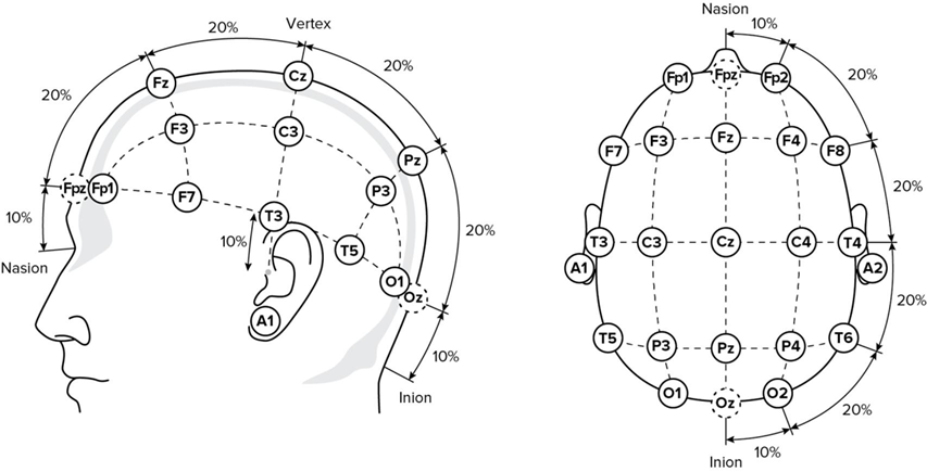
The electrode sites in the 10–20 system have strictly defined designations. Oddnumbered indices correspond to electrodes located above the left hemisphere of the brain, while even-numbered indices correspond to those above the right. Electrodes placed on the earlobes are designated as A1 (left) and A2 (right). If the electrodes are placed on the mastoid, they are designated as M1 (left) and M2 (right).
The electrode locations in the 10–20 system are defined as follows.
- The first line is measured along the sagittal plane from the nasal root (nasion) to the occipital protuberance (inion) and is taken as 100%. The central vertex electrode (Cz) is placed at the midpoint of this line, while the midfrontal (Fz) and central parietal (Pz) electrodes are positioned at distances from Cz equal to 20% of the total length of the line from the nasion to the inion. Measure 10% of this distance from the occipital protuberance towards Cz to mark point Oz. To mark point Fpz, measure 10% of the total distance, but this time from the bridge of the nose towards Cz.
- The second main line (biaricular) runs from the tragus of one ear via the vertex (Cz) to the tragus of the opposite ear. The temporal electrodes (T3, T4) are positioned at a distance of 10% of this line from the tragus. The central electrodes (C3, C4) are positioned to the left and right of Cz at distances equal to 20% of the length of the biauricular line.
- The third line is the line forming half the head circumference. The head circumference is measured such that the line passes through points Fpz, T3, T4 and Oz. The frontal electrodes (Fp1 and Fp2) are placed at a distance of 10% of the head circumference from point Fpz. The occipital electrodes (O1 and O2) are placed at a distance of 10% of the head circumference from point Oz. Points T5, T6, F7 and F8 lie on the circumference line at a distance of 20% of the head circumference from O1, O2, Fp1 and Fp2 respectively.
- Parasagittal lines pass through points Fp1, C3, O1 and Fp2, C4, O2. Frontal (F3, F4) and posterior temporal (T5, T6) electrodes are placed along these lines at equal distances from adjacent points (Fig. 1). In some cases, additional scalp electrodes may be used.
In 2017, the International Federation of Clinical Neurophysiology (IFCN) published amendments to the 10–20 system (Seek M. et al., 2017). According to these amendments:
- T3/T4 leads were renamed T7/T8;
- T5/T6 leads were renamed P7/P8.
A pair of electrodes between which changes in the potential difference are recorded and graphically displayed is called an electroencephalographic lead (Alexandrov M. V. et al., 2018). If both electrodes in the pair are positioned above the brain, such a lead is called bipolar. If the dynamics of the electrical potential are recorded from an electrode positioned above the brain relative to another electrode situated at a distance from the brain tissue (and consequently, the potential change beneath it is practically zero), then such a lead is called a monopolar lead. In this case, the first electrode is considered active, while the second is referred to as the reference electrode. The reference electrode is placed on the earlobe or the mastoid.
In addition to the active and reference electrodes, a ground electrode must be applied. It can be placed on any part of the body but is most commonly attached to the forehead.
Fixation of bridge electrodes for EEG recording
EEG recording is carried out using electrodes that vary in both shape and type of fixation. Bridge electrodes may be used for neurofeedback. Before the exam, the bridge electrodes must be soaked in a 0.9% saline solution for 5–10 minutes. The electrodes are secured under the elastic bands of a special cap. Depending on the size of the patient’s head, the cap is adjusted by tightening or loosening the elastic straps. The electrode placement sites are determined in accordance with the electrode placement system. Prior to placing the electrode, the respective skin area is degreased using a wipe soaked in a 70% isopropyl alcohol solution (hereinafter an “alcohol wipe”). The electrode is connected to the appropriate connector on the device using a lead cable with an alligator clip.
The quality of the EEG recording depends directly on the impedance (skin resistance under the electrode), which should not exceed 40 kΩ(ideally, it should be less than 5 kΩ). If impedance is high, it is recommended to degrease the skin area beneath the electrode once more and moisten the gauze-wrapped surface of the electrode with saline solution using cotton wool or a pipette. In rare cases, the skin area is treated with an abrasive paste. The bridge electrodes are placed on the patient’s head quickly; however, using them does not allow for a prolonged examination (more than 40 minutes), as the electrodes and helmet exert pressure on the patient’s head, causing increased anxiety and additional interference, which undoubtedly has a negative impact on the results of the training. Moreover, the electrodes dry out over time and the impedance increases, which significantly reduces the quality of the recording.
Cup EP electrodes can be used as an alternative to bridge electrodes for EEG biofeedback. The areas of the skin where the electrodes are to be attached are degreased using an alcohol wipe. The electrode is filled with electrode paste and fixed to the degreased skin area. If necessary, the electrodes are additionally fixed using an elastic bandage or adhesive tape. When using cup EP electrodes, the impedance rarely exceeds 10 kΩ; however, if it is higher than the permissible values, the skin under the electrode is treated with an abrasive paste.
1.3. Main Stages of Neurofeedback
1. Psychophysiological and psychological testing prior to the start of BFB therapy.
Prior to treatment, it is recommended to interview the patient to evaluate their personality traits and psycho-emotional state. Sometimes, it is advisable to use testing to quantitatively assess the psychological processes that are supposed to require correction. For example, in patients with attention deficit hyperactivity disorder (ADHD), testing using the Schulte tables or the Schulte–Platonov tables helps to determine baseline levels of concentration, attention switching and mental fatigue. The Beck Depression Inventory (BDI) scale can be used prior to treatment for patients with depression.
The patient should be informed about the training procedure and the duration of treatment. Furthermore, it is important to explain to the patient that the success of BFB therapy depends mainly on their own efforts.
2. Background EEG monitoring prior to the therapy (training) start.
Background EEG monitoring is necessary to establish the baseline threshold values of the controlled parameter.
Electrodes are placed on the patient’s head to record EEG. Electrode impedance is measured. The specialist must ensure that there are no major artifacts during EEG monitoring that could affect the analysis results.
Quantitative EEG characteristics, such as rhythm amplitude, total spectral power of rhythms, ratios of EEG rhythm indices, rhythm asymmetry and others, are used as parameters for EEG biofeedback. Background EEG monitoring allows the determination of individual average values for these parameters. It is best if there is a reference database that allows individual EEG parameter values to be compared with the average values of the corresponding parameters in a control group of the same age. Such a comparison makes it possible to identify statistically significant differences between the patient’s EEG parameters and those of the control group and to use the control group parameters as threshold values for the patient during training sessions. Currently, users of Neurosoft EEG systems can additionally use several validated patient reference databases: NeuroNavigator (ANI NeuroGuide, USA), iSyncBrain (Republic of Korea), BrainMaster (USA) and BrainSys (Russia). However, the use of these reference databases requires additional financial expenditure, which significantly limits their applicability.
If the specialist does not have access to a reference database, the threshold level for the controlled parameter is set based on the baseline EEG values, exceeded by 30% in this case. The threshold value for the controlled parameter may be adjusted during the training session. Background EEG monitoring may only be carried out before the start of the EEG biofeedback course or before each session.
3. Instructing the patient.
At the start of the session, the patient is instructed on how to perform the training.
4. Performing the training.
The training is performed according to the selected BFB protocol. You can select a protocol from the list or create your own protocol.
During the first short cycle (the first sessions of the course), which is aimed at teaching EEG biofeedback, you can help the patient to achieve the state required to fulfill the training conditions, using autogenic techniques or psychological approaches. For example, progressive muscle relaxation, visualisation, deep breathing, etc., are suitable for relaxation-based BFB training. Furthermore, if the patient is unable to meet the set training condition, the specialist can adjust the threshold value of the controlled parameter to make the training easier.
5. Evaluation of the BFB therapy effectiveness.
To evaluate the effectiveness of the therapy, it is necessary to perform background EEG monitoring, as well as a quantitative assessment of the psychological processes requiring correction, and to compare these data with the baseline values obtained prior to the training course.
2. Autonomic BFB Training
When conducting autonomic BFB training, parameters such as EMG amplitude, heart rate based on electrocardiogram (ECG), pulse rate based on a photoplethysmogram (PPG), respiratory rate and other parameters can be used as controlled parameters.
2.1. EMG Biofeedback Training
EMG-BFB training is used in the treatment of motor disorders and for relaxation training (to reduce psychological and emotional stress). Moreover, EMG-BFB training is effective in treating patients suffering from tension-type headaches and pain syndromes, which are primarily associated with dysfunction of muscle activity, most commonly in the form of chronic overexertion.
The choice of electrode placement points depends on the training objective and the type of BFB protocol used (trapezius muscles, frontalis muscles, limb muscles, etc.). A bipolar lead is used for EMG recording; the distance between the electrodes depends on the type of BFB protocol but is most commonly 2–5 cm. To compare data between sessions, the electrode placement points and the distance between them must remain the same throughout the entire course of BFB therapy. It is recommended to position the pair of active electrodes parallel to the main direction of the muscle fibres where electrical activity is measured. In addition to the active electrodes, a ground electrode must be fixed; its position is determined only by convenience of application.
Reusable cup electrodes and disposable adhesive electrodes can be used for EMG recording.
Application of reusable cup electrodes
Degrease the skin at the electrode placement sites with an alcohol wipe. This is particularly important if the patient has oily skin. Fill the electrode cup with conductive adhesive paste, then fix the electrode to the skin. Secure the electrode further with adhesive tape. It is recommended to place the electrodes one at a time immediately after preparing the skin, where possible. In most cases, if the patient’s skin is clean, not flaky and not covered with cream or lotion, no special skin preparation with an abrasive paste is required prior to testing. However, in rare cases, abrasive paste may be used after degreasing the skin to reduce impedance. Then apply the electrode, filled with electrode paste, to the treated skin area and secure it with adhesive tape.
Application of disposable adhesive electrodes
If you use adhesive electrodes, degrease the skin at the electrode placement sites with an alcohol wipe. When the skin is completely dry, apply the electrodes. In rare cases, an abrasive paste may be used to reduce electrode impedance. Sometimes, impedance values may increase during BFB training. To decrease impedance, place a damp cloth or damp cotton wool on the electrodes.
2.2. BFB Based on Cardiovascular System Parameters
Electrocardiographic Bofeedback Training
When conducting ECG-BFB training, a single ECG channel is most commonly recorded. Various types of electrodes may be used for this purpose: clamp ECG electrodes, disposable ECG electrodes with a button clip or reusable cup electrodes.
Application of clamp ECG electrodes
Clamp ECG electrodes with the appropriate cables can be used for ECG recording: the red clamp electrode is placed on the inner (palmar) surface of the lower third of the right forearm and the yellow clamp electrode is placed on the inner (palmar) surface of the lower third of the left forearm. The ground clamp electrode can be positioned on the lower third of the right lower leg, closer to the ankle, or on the upper third of the right or left forearm. Before application, degrease the skin at the electrode placement sites with an alcohol wipe. To improve electrode impedance, it is recommended to apply conductive gel to the contact surface of the electrode.
After the electrodes are applied, connect them using the lead cables to the electronic unit connector, which was configured as an ECG channel for the exam: connect the red electrode cable to the negative input of the channel and the yellow electrode cable to the positive input of the channel. Connect the ground electrode cable to the device connector marked as GRD.
As an alternative to clamp electrodes, peripheral plate electrodes can be used, which are fixed using a special band.
Application of disposable ECG electrodes
When using disposable ECG electrodes with a button clip, they should be placed on the patient’s torso using a modified standard lead II. The skin at the electrode placement sites should first be degreased using an alcohol wipe. No additional application of conductive gel is required when using disposable ECG electrodes. When the electrodes are placed, connect them using the lead cables to the electronic unit connector, which was configured as an ECG channel for the exam: the cable of the electrode located in the right supraclavicular region to the negative input of the channel and the cable of the electrode located in the left hypochondrium to the positive input of the channel. The ground electrode can be placed in the left subclavian region and connected to the device connector marked GRD using a special cable.
As an alternative to disposable ECG electrodes, reusable cup electrodes can be used, which are placed on the patient’s torso using a modified standard lead II. Application of reusable cup electrodes is described above.
Photoplethysmography-based BFB Training
It is possible to conduct training based on the pulse rate calculated using a photoplethysmogram (PPG). PPG is recorded using a special sensor connected to a pulse oximeter.
A reusable or disposable SpO2 sensor is attached to the patient’s finger (Fig. 2). If necessary, the sensor cable can be fixed to the back of the hand using adhesive tape. The SpO2 sensor is connected to a separate SpO2 unit, which is plugged into a computer’s USB port. For the Neuron-Spectrum-4EPM/S, Neuron-Spectrum-5/S and Neuron-Spectrum-AM systems, the SpO2 sensor must be connected to the appropriate electronic unit connector.
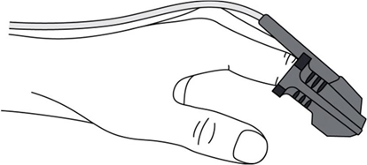
Please note that nail polish on the patient’s finger may affect pulse rate measurement. Make sure to remove nail polish from the finger on which the sensor is placed.
2.3. BFB Training Based on Respiratory Parameters
Proper breathing is the basis of any relaxation technique, which is extremely important for relieving psychological and emotional tension and for overall health improvement.
Breathing movements are accompanied by changes in the volume of the thoracic and abdominal cavities. This phenomenon is used to record respiratory effort during BFB training sessions. An inductive belt with a sensor is recommended for recording respiratory effort. A single belt is enough for calculating respiratory rate; however, when assessing chest and abdominal movement in relation to the overall breathing pattern, two belts must be used. One belt is fixed at the level of the axillary fossae (to record chest movements), the other just above the iliac crests (to record abdominal movements).
2.4. Galvanic Skin Response
The galvanic skin response (GSR) is one of the most sensitive methods for the dynamic monitoring of a person’s psychophysiological state.
The placement of the recording electrodes on the palm is shown in Fig. 3. Place the first recording electrode on the second phalanx of the middle finger. Connect the cable of this electrode to the + input of the polygraphic channel. Fix the second recording electrode on the palm, in line with the second interdigital space, at a distance of 3 cm from the skin fold located at the level of the metacarpophalangeal joints. Connect the electrode cable to the − input of the polygraphic channel. Position the ground electrode at a convenient place. Depending on the training objectives and the type of BFB protocol used, the position of the electrodes may be adjusted.
Both disposable adhesive electrodes and reusable cup EP electrodes can be used to record the GSR.
When preparing the skin, avoid applying excessive mechanical pressure, as this may affect the sweat glands, the response of which is directly recorded. Before applying the electrodes, degrease the patient’s skin using an alcohol wipe. When the skin is dry, apply a disposable adhesive electrode to the degreased area. For a reusable electrode, fill the electrode cup with electrode paste and apply the electrode to the degreased skin, fixing it additionally with adhesive tape.
Factors affecting GSR recording:
- high electrode impedance;
- air temperature in the room does not meet standard operating conditions;
- stress or physical activity prior to the exam;
- increased patient’s temperature;
- external distractions (noise, loud sounds); · use of medications with a vegetotropic effect;
- patients aged over 65.
2.5. Stages of BFB Therapy
BFB therapy, based on parameters regulated by the autonomic nervous system, is carried out following the same stages of neurofeedback training (EEG-BFB).
1. Introduction to training.
Before starting therapy, it is recommended to explain the training procedure and the duration of treatment to the patient. It is also important to explain to the patient that the success of the therapy depends primarily on their effort.
2. Background monitoring of physiological signals prior to the training start.
Background monitoring (EMG, respiratory, etc.) is necessary to establish threshold values for the controlled parameter during training. Position the sensors or electrodes according to the montage and BFB protocol. The position of the active electrodes and the distance between them must remain the same both during background monitoring and throughout the training sessions. Check the impedance; it must be within the green range. During physiological signal monitoring, ensure that there are no artifacts that could affect the analysis results. Quantitative characteristics such as EMG amplitude, respiratory rate, heart rate, pulse rate and GSR amplitude can be used as BFB parameters.
3. Instructing the patient.
The patient is instructed on how to perform the training. Before the relaxation sessions, the patient may be introduced to certain relaxation techniques and psychophysiological approaches that facilitate quick learning and enhance the effectiveness of BFB therapy.
4. Performing the training.
The training is performed according to the selected BFB protocol. You can select a protocol from the list or create your own protocol.
5. Evaluation of the BFB therapy effectiveness.
3. Application of Biofeedback
The Neurosoft equipment enables the use of many well-known BFB protocols. The company does not develop biofeedback protocols, nor does it participate in research demonstrating their clinical efficacy.
This section provides a brief description of several well-known BFB protocols reviewed in the scientific literature. When selecting a protocol for BFB therapy, it is recommended that you refer to the full description via the provided reference.
3.1. Attention Deficit Hyperactivity Disorder
According to various estimates, 3–7% of schoolchildren suffer from attention deficit hyperactivity disorder (ADHD). Moreover, in 40–60% of patients with this diagnosis, ADHD symptoms persist beyond the age of 18. Clinical guidelines for the management of children with ADHD recommend a multimodal approach, including non-pharmacological methods such as neurofeedback. A number of studies on quantitative EEG report an increase in the spectral power of slow-wave activity in patients with ADHD compared to the norm (Kropotov J. D., 2010). As absolute spectral power indices vary considerably, a relative parameter was proposed for use: the ratio of theta wave power (4–8 Hz) to beta wave power (13–21 Hz) (Kropotov J. D., 2010). This parameter was called the “inattention index” (Monastra V. J. et al., 1999). In 6–7-year-old children with ADHD, the theta/beta ratio was three times higher than normal (Kropotov J. D., 2010). It is assumed that normalisation of this parameter will contribute to an improvement of the patient’s condition.
There are several neurofeedback protocols used in the treatment of children with ADHD. Their effectiveness is evaluated in scientific reviews (Arns M. et al., 2014) and meta-analyses (Van Doren J. et al., 2019).
3.1.1. EEG Biofeedback (Beta Protocol) for the ADHD Treatment (Stark et al., 2000b)
Stages of BFB therapy
1. Assessment of the patient’s psychological and emotional state.
Schulte tables are used to assess attention span, adaptability and mental fatigue.
2. Background EEG monitoring.
At the start of the neurofeedback course, background EEG is recorded for 5–10 minutes. EEG parameters are assessed in the same leads used for training.
Subsequently, EEG monitoring is carried out no more than once a week.
3. Pre-training.
When hyperactivity is severe and interferes with BFB training, EMG or SMR training is carried out prior to the main training.
· SMR training (a component of the treatment program for patients with severe hyperactivity).
Training conditions: increasing the power of the sensorimotor rhythm spectrum.
Montage: C1–C5, ground electrode.
Number of sessions: 20–30 sessions.
· EMG training (a component of the treatment program for patients with severe hyperactivity).
Training conditions: decreasing EMG amplitude.
Recommended leads: two electrodes are placed on the frontalis muscles; the ground electrode may be placed on the mastoid.
4. Beta stimulation training (the main phase of therapy).
Training conditions: the authors of this BFB protocol recommend two options for EEG training:
· increasing spectral power in the beta band;
· decreasing the ratio of spectral power levels in the theta and beta bands.
Montage: FCzCPz (the FCz point is located halfway between Cz and Fz; the CPz point is located halfway between Cz and Pz), ground electrode.
Session duration: training duration — 3–5 minutes at the start of treatment, with the duration subsequently increased to 15 minutes; number of trainings — 4–5 during the session; total session duration — at least 20 minutes.
Number and frequency of sessions: 30–40 (sometimes 60) sessions; 2–3 times a week.
5. Evaluation of the BFB therapy effectiveness.
Follow-up monitoring for 4 months.
3.1.2. Theta/Beta Protocol (Gevensleben et al., 2009, 2010; Gelade et al., 2017; Duric et al., 2012*)
Stages of BFB therapy
1. Background monitoring of EEG activity.
Background EEG is performed for 5 minutes at the beginning and end of the session to determine the threshold values of the controlled parameter and to assess the dynamics of changes in the parameter.
2. Preparatory EMG training* (a component of the treatment program for patients with severe hyperactivity) (Duric N. S. et al., 2012).
Training conditions: decreasing EMG amplitude.
Recommended leads: two electrodes are placed on the frontalis muscles; the ground electrode must also be applied.
3. Theta (4–8 Hz)/beta (13–20 Hz) training (main training).
Training conditions: decrease in the ratio of activity in the theta band (4–8 Hz) to activity in the beta band (13–20 Hz).
Montage: CzA, ground electrode.
Session duration: training duration — from 5 (at the start of treatment) to 10 minutes; number of trainings — 5–6 during the session; total session duration — at least 40–50 minutes.
Number and frequency of sessions: 30–36 sessions; 2–3 times a week.
Follow-up monitoring for 4 months.
3.1.3. Theta↓, Beta↑ or Theta (4–8 Hz)/Beta (13–20 Hz)↓ Protocol (Meisel et al., 2014)
Stages of BFB therapy
1. Psychological assessment.
2. Background EEG monitoring.
At the start of each session, background EEG monitoring is performed for 30 seconds. The background values of the controlled parameters are set as threshold values for training.
3. Training.
Training conditions: decreasing activity in the theta band (4–8 Hz), increasing activity in the beta band (13–20 Hz) and decreasing the ratio of activity in the theta band (4–8 Hz) to activity in the beta band (13–20 Hz).
Montage: CzA (children), FCzA (adults), ground electrode.
Session duration: 6 trainings for 4 minutes each, total duration — 35 minutes.
Number and frequency of sessions: 40 sessions; 2 times a week.
4. Psychological assessment (evaluation of treatment effectiveness).
Follow-up monitoring for 2–6 months.
3.1.4. Theta/SMR Protocol (Steiner et al., 2014).
Stages of BFB therapy
1. Training.
Training conditions: decreasing the ratio of EEG activity in the theta band to activity in the sensorimotor rhythm band (12–15 Hz): theta (4–8 Hz)/SMR (12–15 Hz)↓.
Montage: CzA, ground electrode.
Session duration: 45 minutes.
Number and frequency of sessions: 40 sessions; 3 times a week.
Follow-up monitoring for 6 months.
3.2. Addictive Disorders
Neurofeedback is a universal component of rehabilitation programs for patients with almost any addictive disorder. BFB training, conducted by a qualified specialist, directly influences the universal central mechanisms involved in the development of addiction; it is, therefore, a pathogenetic method of treating addictive conditions (Stark M. B. et al., 2000a).
3.2.1. EEG Biofeedback (Alpha-Training) for the Treatment and Rehabilitation of Addictive Disorders (Stark et al., 2000a)
Stages of BFB therapy
1. Psychological assessment.
Interview with patient and evaluation of psychoemotional state (Lüscher color test, Beck Depression Inventory (BDI), Hamilton Rating Scale for Depression and others).
2. Temperature-myographic training (can be included into the program at the beginning of treatment or at the beginning of each session).
Training conditions: increasing temperature, decreasing EMG amplitude.
3. Background EEG monitoring.
At the beginning of the neurofeedback session, background EEG activity is recorded, which helps to determine not only the threshold values of the controlled parameters but also the positions of the active EEG electrodes. If alpha activity prevails in the left frontal lobe or if there is no interhemispheric asymmetry, the authors of the protocol recommend using the F4C4 lead for training. If right-sided alpha activity prevails, using the F3C3 lead is recommended for training.
4. Theta stimulation training (at the beginning of the treatment course or at the beginning of each session).
Training conditions: increasing spectral power in the theta band.
Montage: F4C4 or F3C3, ground electrode.
5. Alpha stimulation training.
Training conditions: increasing spectral power in the alpha band.
Montage: F4C4 or F3C3, ground electrode.
Session duration: 30 minutes.
Number of sessions: 20–30 sessions.
Follow-up monitoring for 2–6 months.
Contraindications for therapy
· endogenous mental disorders;
· intellectual disability, stage three alcohol or drug addiction;
· the protocol is ineffective when used in conjunction with intermittent medication.
3.2.2. Peniston-Kulkosky Neurofeedback Therapeutic Protocol for Patients with Chronic Alcoholism (Peniston, Kulkosky, 1999)
Stages of BFB therapy
1. Psychological assessment.
Interview with patient, evaluation of psychoemotional state (Lüscher color test, Beck Depression Inventory (BDI), Minnesota Multiphasic Personality Inventory (MMPI) and others).
2. Background multichannel EEG monitoring.
Before the start and after the completion of neurotherapy, a 16-channel EEG recording is performed, during which the individual alpha rhythm peak (in Hz), the average alpha rhythm amplitude and the alpha rhythm index are assessed.
3. Temperature BFB training combined with autogenic training.
Training conditions: increasing temperature.
4. Background one-channel EEG monitoring.
Background EEG monitoring consists of 2 tests:
· EEG monitoring of a patient with eyes closed for 5 minutes, during which the amplitude of the alpha rhythm and the alpha and theta rhythm indices are assessed;
· EEG monitoring of a patient with eyes open, during which the patient is asked to focus on any object in the room for 5 minutes. When the beta rhythm index reaches 40–50%, the amplitude (index) of the beta rhythm is assessed.
5. Alpha-theta stimulation training.
Training conditions: increasing spectral power (or index) in both the alpha and theta bands (the training is performed with eyes closed).
Montage: O1A1, ground electrode (А2).
Session duration: 30 minutes.
Number and frequency of sessions: 30 sessions; within 28 days.
6. One-channel monitoring (O1A1).
Monitoring of EEG activity at the end of each alpha-theta stimulation training session, based on which the average amplitude of the alpha rhythm and the indices of the beta, alpha and theta rhythms are assessed.
A 13-month follow-up monitoring indicates no relapses in the group of patients with chronic alcoholism who completed a full course of alpha-theta training.
3.3. Tension-Type Headache
Tension-type headache (TTH) is a bilateral, diffuse headache characterized by a tightening or pressing sensation of mild to moderate intensity. It is one of the most common types of headache. The prevalence of chronic TTH is approximately 2–3% of the general population. The primary cause of TTH is considered to be muscle tension, which leads to the development of myotonic syndrome — painful tension in the muscles surrounding the head and neck. In addition, emotional stress can intensify muscle tension, which ultimately leads to TTH. Combined therapy using both pharmacological and non-pharmacological methods is highly effective in treating chronic TTH (Filatova E. G, 2005).
3.3.1. BFB protocol for the treatment of patients with TTH: decreasing EMG activity of the trapezius and frontalis muscles (Mullally et al., 2009).
Training conditions: decreasing EMG amplitude.
Montage: trapezius and frontalis muscles.
Session duration: 50 minutes.
Number of sessions: 10.
3.4. Post-Traumatic Stress Disorder
Post-traumatic stress disorder (PTSD) is a mental disorder that develops as a result of a powerful traumatic event of a threatening or catastrophic nature and is accompanied by extreme stress. The main clinical manifestations include the re-experiencing of the traumatic event in the present in the form of vivid intrusive memories, flashbacks or nightmares, which are often accompanied by anxiety and panic, but may also include anger, resentment, guilt or hopelessness and a desire to avoid internal and external stimuli that remind the individual of the stressor or are associated with it. According to the Clinical Guidelines of the Russian Society of Psychiatrists “Post-Traumatic Stress Disorder” (2023), patients with PTSD are recommended to undergo BFB therapy aimed at training the brain to self-regulate without the use of external stimulation (strength of recommendation — A, level of evidence — 1).
The use of a non-invasive form of neurofeedback (BFB therapy) is recommended, regardless of the type of neuroimaging (using EEG and real-time functional magnetic resonance imaging), with the aim of training the brain to self-regulate without the use of external stimulation. Therapy is conducted daily. The duration of each session is 15–20 minutes and the number of sessions per course is 10–12 (Clinical Guidelines “Post-Traumatic Stress Disorder,” 2023).
3.4.1. BFB protocol for rehabilitation of patients with chronic PTSD (van der Kolk, 2016)
Stages of BFB therapy
1. Psychological assessment (Clinical-Administered PTSD Scale, CAPS).
2. Background EEG monitoring.
Once the background EEG activity stabilizes, the thresholds for the target training parameters are calculated:
· the spectral power threshold in the 2–6 Hz range is set so that it is exceeded in the background 35% of the time during the background test;
· the spectral power threshold in the 22–36 Hz range is set so that it is exceeded in the background 25% of the time during the background test;
· the spectral power threshold in the 10–13 Hz range is set so that it is exceeded in the background 65% of the time during the background test (in some cases, the 10–13 Hz range is adjusted based on the patient’s symptoms).
3. Neurofeedback.
Training conditions: decreasing spectral power in the 2–6 Hz and 22–36 Hz bands, accompanied by an increase in spectral power in the 10–13 Hz band.
Montage: T4P4, ground electrode (A1).
Feedback: video games, music.
Session duration: training begins with a 12-minute session; thereafter, the session duration gradually increases by 3 minutes if the task is successfully completed; the maximum session duration is 30 minutes.
Number and frequency of sessions: 24–27 sessions, 2 times a week.
There is also evidence of the successful use of the Peniston–Kulkosky protocol for the treatment of patients with PTSD (Nicholson A. A. et al., 2020).
3.5. Insomnia
Insomnia is a clinical syndrome characterized by complaints of disturbed nighttime sleep (difficulty falling asleep, staying asleep or waking up earlier than desired) and associated disturbances during daytime wakefulness, occurring even when there is sufficient time and opportunity for sleep. Based on this definition, the prevalence of insomnia in the general population is estimated at 9–15% (Poluektov M. G., 2016).
3.5.1. SMR/theta stimulation protocol (Białkowska J., 2022)
Stages of BFB therapy
1. Psychological assessment (Pittsburgh Sleep Quality Index (PSQI), Beck Depression Inventory (BDI), State-Trait Anxiety Inventory (STAI)).
2. Background EEG monitoring.
3. SMR/theta stimulation training.
Training conditions: increasing the amplitude of the SMR rhythm (12–15 Hz) and simultaneously decreasing the amplitude of the theta rhythm (4–7 Hz).
Montage: C4А1, ground electrode.
Session duration: 10 three-minute training sessions, with a total duration of 30 minutes.
Feedback: video and/or audio.
Instructions for the patient: perform the relaxation exercises while following the audiovisual cues.
Number of sessions: 30.
4. Psychological assessment (Pittsburgh Sleep Quality Index (PSQI), Beck Depression Inventory (BDI), State-Trait Anxiety Inventory (STAI)).
Additionally, the SMR stimulation protocol is also used to treat patients with insomnia (Hoedlmoser K. et al., 2008).
References
References
1. Aleksandrov MV, et al. Electroencephalography. Saint Petersburg. Strategy of the Future [Strategia budushchego]. 2018. (In Russ.)
2. Clinical Guidelines “Post-Traumatic Stress Disorder” (approved by the Ministry of Health of the Russian Federation). 2023. (In Russ.)
3. Kropotov JD. Quantitative EEG, cognitive evoked potentials of the human brain and neurotherapy. Donetsk. Zaslavsky. 2010:299-301. (In Russ.)
4. Poluektov MG, et al. Project of clinical recommendations on diagnosis and treatment of chronic insomnia in adults. Consilium Medicum. Neurology and Rheumatology. 2016;2(Suppl):41-51. (In Russ.)
5. Polyaev BA, et al. Safety and efficacy of the neurofeedback course on a protocol of strengthening of the spectral power of the alpha rhythm in top-class athletes: placebo-controlled study. Sports Medicine: Research and Practice. 2013;4:34. (In Russ.)
6. Filatova EG. Tension-type headache. Handbook for practitioners doctors. 2005;4:57-60. (In Russ.)
7. Stark MB, et al. Electroencephalographic biofeedback (alpha-theta training) for the treatment and rehabilitation of addictive disorders (pathological addictions) and depression. Methodological guidelines of the Ministry of Health of the Russian Federation No. 99/174, December 28, 2000. Novosibirsk. 2000a:34. (In Russ.)
8. Stark MB, et al. Electroencephalographic beta-stimulation training for the treatment of attention deficit disorder and associated conditions. Methodological guidelines of the Ministry of Health of the Russian Federation No. 99/175, December 29, 2000. Novosibirsk. 2000b:15. (In Russ.)
9. Arns M, Heinrich H, Strehl U. Evaluation of neurofeedback in ADHD: the long and winding road. Biol Psychol. 2014;95:108-115.
10. Białkowska J, Mroczkowska D, Boraczyński M. Subjective Improvement of Sleep in Insomnia Patients Treated at a Day Rehabilitation Centre After the Use of EEG Neurofeedback Therapy–A Pilot Study. Medical Rehabilitation, 2023;27(1):27-32.
11. Duric NS, Assmus J, Gundersen D, Elgen IB. Neurofeedback for the treatment of children and adolescents with ADHD: a randomized and controlled clinical trial using parental reports. BMC Psychiatry. 2012;12:107.
12. Geladé K, Bink M, Janssen TW, van Mourik R, Maras A, Oosterlaan J. An RCT into the effects of neurofeedback on neurocognitive functioning compared to stimulant medication and physical activity in children with ADHD. Eur Child Adolesc Psychiatry. 2017;26(4):457-468.
13. Gevensleben H, Holl B, Albrecht B, et al. Is neurofeedback an efficacioustreatment for ADHD? A randomised controlled clinical trial. J Child Psychol Psychiatry. 2009;50(7):780-789.
14. Gevensleben H, Holl B, Albrecht B, et al. Neurofeedback training in children withADHD: 6-month follow-up of a randomised controlled trial. Eur Child Adolesc Psychiatry. 2010;19(9):715-724.
15. Gilbert C, Moss D. Biofeedback and biological monitoring. Handbook of mindbody medicine in primary care. 2003:109-122.
16. Hoedlmoser K, Pecherstorfer T, Gruber G, et al. Instrumental conditioning ofhuman sensorimotor rhythm (12-15 Hz) and its impact on sleep as well as declarative learning. Sleep. 2008;31(10):1401-1408.
17. Jasper HH. Ten-twenty electrode system of the international federation.Electroencephalogr Clin Neurophysiol. 1958;10:371–375.
18. Meisel V, Servera M, Garcia-Banda G, Cardo E, Moreno I. Reprint of“Neurofeedback and standard pharmacological intervention in ADHD: a randomized controlled trial with six-month follow-up”. Biol Psychol. 2014;95:116125.
19. Monastra VJ, Lubar JF, Linden M, et al. Assessing attention deficit hyperactivitydisorder via quantitative electroencephalography: an initial validation study. Neuropsychology. 1999;13(3):424-433.
20. Mullally WJ, Hall K, Goldstein R. Efficacy of biofeedback in the treatment ofmigraine and tension type headaches. Pain Physician. 2009;12(6):1005-1011.
21. Nicholson AA, Tomas R, Rakesh J, Ruth AL. Regulating posttraumatic stressdisorder symptoms with neurofeedback: Regaining control of the mind. Journal of Military, Veteran and Family Health. 2020;6(Suppl 1):3-15.
22. Peniston EG, Kulkosky PJ. Neurofeedback in the treatment of addictivedisorders. Introduction to quantitative EEG and neurofeedback. 1999:157-179.
23. Seeck M, Koessler L, Bast T, et al. The standardized EEG electrode array of theIFCN. Clin Neurophysiol. 2017;128(10):2070-2077.
24. Steiner NJ, Frenette EC, Rene KM, Brennan RT, Perrin EC. Neurofeedback andcognitive attention training for children with attention-deficit hyperactivity disorder in schools. J Dev Behav Pediatr. 2014;35(1):18-27.
25. van der Kolk BA, Hodgdon H, Gapen M, et al. A Randomized Controlled Study ofNeurofeedback for Chronic PTSD. PLoS One. 2016;11(12):e0166752.
26. Van Doren J, Arns M, Heinrich H, Vollebregt MA, Strehl U, K Loo S. Sustainedeffects of neurofeedback in ADHD: a systematic review and meta-analysis. Eur Child Adolesc Psychiatry. 2019;28(3):293-305.