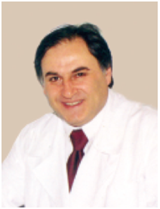
Khidirbegishvili O.E.
Dentist. Tbilisi, Georgia
Abstract. The article presents paradoxical situations in modern cariesology and discusses methods of their elimination. Special attention is paid to the scientific validity of the classifications used, including the WHO classification..The disadvantages of these classifications are described in detail, related to the imperfection of diagnostic methods, the variety of forms and clinical manifestations of caries, which makes it difficult to determine the resistant status of carious pathology.At the same time, the principles of constructing modern clinical classifications of caries are formulated.
Keywords: dental caries,fissure caries,isolated and combined carious cavities,superficial caries, caries arrested.
INTRODUCTION
Today, cariesology has reached a sufficient level of development to provide clinicians with adequate knowledge in the field of etiology, clinic and treatment of caries.However, strangely enough, there are still many paradoxical situations and scientific misconceptions in cariesology that have not yet been eliminated.
I will say more, sometimes it seems that mass psychosis reigns in dental science in general, otherwise how can we explain that for decades we have been misinterpreting some generally accepted postulates and rules of diagnosis, we cannot definitively decide the validity of the application of a particular technique, etc..That is why clinicians still often make clinically unjustified diagnoses. However, what is especially striking is that such a situation is absolutely not confused by international dental organizations, and many well-known clinicians, it would seem, have already come to terms with such an inadequate situation!
MATERIALS AND METHODS
Let's consider the existing paradoxical situations, starting with one of the oldest of them, when the outstanding American scientist Green Vardiman Black, the son of a peasant woman and a cabinetmaker, who actually did not have not only higher medical education, but also a full–fledged secondary education (only at the age of 42 he became a certified doctor, having independently passed exams), proposed in 1881 a classification of the localization of carious cavities[1].
Curiously, Black proposed his classification for the use of inlays, fillings made of gold and amalgam, the use of which involved the removal of not only carious, but also a significant number of unaffected tooth tissues to ensure reliable fixation of the seal.Therefore, at that time, its classification met the requirements of clinicians, but after the death of the scientist, any filling materials that appeared on the dental market unreasonably sought to adapt to its classification.Unfortunately, the proponents of such a position were absolutely not embarrassed, that Black's surgical approach was designed only for the use of gold and amalgam fillings[1].
Even composites (1962) and glass ionomers (1970), which finally made it possible to carry out techniques of minimal invasive intervention (an alternative to the Black method), apparently as a sign of gratitude to an outstanding scientist, oddly enough, were adapted to this classification.
Having carried out a scientific analysis of the Black classification used today for the use of modern sealing materials, it is possible to find a number of significant shortcomings in it.For example, it is difficult to agree with the interpretation of Class V, because if the cervical region is located around the neck of the tooth as a whole, then caries in this area on the contact surfaces should also be considered cervical[2].
This is once again evidenced by the isolation of circular caries by the Australian scientist Graham Mount as a type of cervical caries [3].It is also important that when an adjacent tooth is missing, the open contact surface is no longer a cariesogenic zone and the primary carious process in this area can occur only in the cervical cariesogenic zone (cervical caries).
Graham Mount's tactics also deserve attention, according to which he attributes Class VI cavities, depressions in the area of Carabelli tubercles, etc. to fissure caries[3].I consider this approach quite justified, since class VI lesions, which were later added to Black's classification by other authors, are rarely diagnosed in the clinic and occur only when there are depressions (fissures, pits, grooves, etc.) on the tops of the tubercles of the lateral and cutting edges of the front teeth [2].
A significant disadvantage of Black's classification is the diagnosis of only progressive forms of carious lesions, without taking into account the early signs of carious lesions.This approach thereby limits the possibility of evaluating the effectiveness of preventive interventions in the early stages of caries and underestimates the prevalence and severity of the disease[2].
In addition, the classification does not pay attention to the localization of the carious process on the exposed surface of the root (root caries), which undoubtedly reduces the quality of diagnosis. In addition, six classes of carious cavities according to Black cannot cover the full variety of variants of damage to hard tooth tissues found in the clinic, therefore, cavities that do not fit into this classification are classified as atypical[4].The identification of such cavities once again indicates the limited scope of application of Black's classification.
At the same time, it is important to note that the tactics of diagnosis and treatment of carious lesions are clearly influenced not only by their localization in cariesogenic zones, but also by belonging to isolated or combined cavities. As is known, isolated cavities are located on one or more tooth surfaces that do not connect to each other.And when several carious cavities on different surfaces of the tooth connect to each other, then we are talking about combined cavities [2].Such a separation of cavities is essential for the tactics of formation of a carious cavity, since in combined cavities, after preparation, the main and additional sites are often isolated.
Paradoxically, in Black's classification, only class IV refers to combined lesions of the frontal teeth, and all the others are isolated. But combined lesions of the contact surfaces of the lateral teeth in violation, for example, of the integrity of the chewing surface, due to more difficult surgical access, significant occlusal loads and a more complex configuration of the contact point, have their own characteristics of treatment and restoration of such lesions[2].
That is why, in the classification of the localization of carious cavities, there should definitely be a strict differentiation of cavities into isolated ones (fissure, contact and cervical caries)and combined.Moreover, I propose to classify combined carious cavities as follows:
l. Carious cavities on the chewing or cutting surface with a diverse combination of adjacent surfaces involved;
2. Carious cavities on the proximal surfaces with a diverse combination of involved lingual and labial surfaces;
3. Extensive carious cavities with the absence of one or more tooth surfaces.
Only such a tactic of distinguishing three classes of combined cavities allows us to take into account the full variety of combined lesions[2].
However, the most disastrous and traumatic in relation to tooth tissues turned out to be Black's surgical approach, designed on the principle of preparation "expansion for prevention", which is based on the method of preventive expansion of carious cavities to the so-called cariesimmune zones.The method involves wide excision of caries susceptible areas (fissures, contact surfaces, maxillary area) to caries-immune areas (cusps, equator, crown roundings) with the creation of a box-shaped cavity[1].
According to Black and his followers, such dissection tactics allegedly prevented the occurrence of recurrent caries due to the deliberate expansion of the boundaries of the carious cavity in the cariesogenic zone to the cariesimmune zones[1].That is why the cavity design was classified, standardized, and healthy dental tissue was sacrificed to geometric perfection in order to supposedly prevent possible complications.
As a result, a standard rather traumatic dissection system was formed, when even small foci of demineralization required the removal of significant amounts of healthy dental tissue. At the same time, the edges of the cavity were formed within the zones of natural self-cleaning, which, according to Black's erroneous opinion, prevented the resumption of the carious process in the sealed tooth[2].
Unfortunately, at that time Black did not know about the main scourge of filling materials – polymerization shrinkage, which occurs after their solidification and as a result of which cracks form between the tooth tissues and the filling material[5].It is in these polymerization crevices in the caries-immune regions that microorganisms multiply, forming recurrent caries (Fig.1).Thus, the use of the Black dissection technique did not save from the occurrence of recurrent caries on caries-immune surfaces, but, on the contrary, further contributed to the occurrence of recurrent caries (see below) [2].
Fig. 1. Consequences of polymerization shrinkage.
In this case, the principle of extending the boundaries of preparation according to Black to the zones of natural self-purification does not prevent the reproduction and colonization of bacteria in these microspaces, since the bacterial plaque is not on the smooth surface of the caries-immune zone, but in artificial depressions resulting from polymerization shrinkage of the material, where its mechanical purification is difficult[2].
That is why modern science has proven that the main reason for the resumption of the carious process at the boundaries of the supplied seal is polymerization shrinkage of the material, which is much more difficult to combat, since none of the existing methods of combating polymerization shrinkage provides a full guarantee of preventing this complication[5].
In addition, it has been proven that the larger the size of the cavity and, accordingly, the volume of the filling material introduced into it, the greater the strength and negative consequences of polymerization shrinkage.Therefore, the removal of intact tissues by Black to expand the boundaries of the cavity to the immune zones not only makes no sense, but also causes an even greater likelihood of complications.
I would also like to note that the principle of "expansion for prevention" no longer corresponds to an infectious disease, but to an oncological disease, in the treatment of which healthy tissues are excised to prevent metastases[2].
In addition, Black did not properly appreciate the most important discovery of that time, made by the father of preventive dentistry Miller, who in the book "Microorganisms in the human oral cavity" (1890), in fact, proved the infectious nature of carious pathology[6].
Having properly evaluated this discovery, it can be stated that an infectious disease like caries cannot be completely cured only surgically.
Black, on the other hand, believed that the only effective method of eliminating carious lesions was the removal of a significant amount of healthy tissue along with the affected ones, followed by reconstructive therapy. That is why Black proposed a surgical approach to the treatment of caries, which, in the absence of a proper scientific understanding of the formation of a carious lesion at that time, was in principle quite logical, but not today, when the pathogenesis of caries has been fully studied[1].
I will say more, it is precisely because of the destructive method of Black in the absence of effective painkillers and turbine drills, due to the special traumatism and painfulness of this method, visiting the dentist has become a living hell for the patient. That is why I would not like to be in the role of a patient who, for some reason, diligently expands the boundaries of an insignificant carious cavity without anesthesia to cariesimmune zones using foot drills[2].
To a large extent, because of Black's dissection technique, there was a general fear of visiting the dentist.
However, despite this, even today, in the era of minimal invasive intervention, modern textbooks on therapeutic dentistry still suggest the use of the destructive principle of Black's dissection. I will say more, even seemingly well-known and experienced clinicians still promote the traumatic and destructive surgical method of Black's dissection for tooth tissues.
In this regard, it is surprising, and sometimes shocking, that Prof Domenico Ricucci described in his book "Endodontics. Clinical and Biological Aspects", a rather strange experiment with the carious tooth in Fig. 3-2, which was subsequently extracted ostensibly for orthopaedic reasons as a result of enlargement of the dental alveolar process.
Figure 3-2 from Domenico Ricucci's book.
In order to prove the presence of microorganisms in dense pigmented dentin in this experiment, Mr. Riccucci prepared a shallow carious lesion in a fissure on the second molar of the upper jaw using the rather traumatic Black method, deliberately leaving a dense pigmented layer of dentin at the bottom of the cavity. After the tooth was removed, he prepared preparations for histological examination.
Not only that, diagnosing the initial lesion, he barbaric method, using Black's methodology, prepares the tooth, so the famous scientist apparently forgot that it has long been proved that the presence of microorganisms in pigmented dense dentin, which with timely adequate treatment is not dangerous under the restoration, as a result of which doctors around the world in the treatment of such lesions never remove dense pigmented dentin.
Anyway, first of all, I just feel sorry for this removed tooth, which, even because of the dental alveolar extension, could certainly have been easily saved (at least preserve healthy sealed roots), and not removed for the sake of this useless virtual experiment.
It has come to the point that with non-carious lesions, some "advanced" clinicians, paradoxically, also use Black's dissection tactics. I will say more, in the textbook on therapeutic dentistry edited by E.V. Borovsky (2003), it is recommended to use this Black technique when treating teeth with non-carious lesions [4]. It is unlikely that the legendary scientist, who proposed his own dissection technique for carious lesions, would agree with such an inadequate use of it.
The fanatical faith of many clinicians in Black's surgical approach could not be shaken even by the method of ‘biological expediency’ proposed in 1948 by I.G. Lukomsky, according to which only caries-affected tissues are excised [8].And what is particularly unacceptable, to this day it is still not actually accepted to question Black's destructive approach of preparation, which is harmful to the tooth tissues, and it is still insistently proposed for use in the clinic. Black foresaw that his principles and ideas would one day be challenged and that scientists would invent new, more effective ways of treating teeth[1].
That's why he liked to repeat that "the main rule of a professional is to constantly learn new things". Unfortunately, many of today's "advanced" scientists who do not respond to my messages and refuse to publish my articles, ignoring my attempts to correct paradoxical situations in dentistry, do not adhere to the precepts of the great scientist at all.
For example, the editor-in-chief of the American journal JADA, Tim Wright, refused to publish my article in which I proposed to modernize the modern classification of caries ICDAS, explaining it in fact as follows :"Of course, the classification is not without drawbacks, but American doctors are used to it and therefore there is no point in modernizing it".There is nothing to say – the "wise" and "philosophical" logic of the reason for refusal!
Clearly, the classification and methodology of Black's dissection is incompatible with the modern philosophy of minimal intervention and prevention of caries. Undoubtedly, the era of Black, which has systematically stirred the consciousness of clinicians around the world for more than a century, has become a thing of the past and a new era has begun – the era of minimal invasive intervention.
So today the famous Green Black slogan "Extension for Prevention" needs to be reworded to "Prevention of Extension".
At the same time, it should be noted that, of course, even today it is sometimes necessary to minimally remove healthy tooth tissues, but not to prevent recurrent caries, but mainly for reliable fixation of restoration. In addition, if, after the removal of carious tissues, the prepared cavity comes into contact with a deep fissure, which is not affected by caries, then in this case it is advisable to open it and seal it together with the main cavity.This prevents not recurrent caries, but the possibility of caries in an unaffected fissure. Agree, these are completely different concepts[2].
Paradoxically, significant disadvantages are characteristic not only of the Black classification, but also of other used clinical classifications of caries, as a result of which it is sometimes impossible to make a reasonable diagnosis of caries. I will say more, considering the classifications used in different countries, it is amazing how different and contradictory they are, and sometimes it even seems that we live on different planets.
Unfortunately, there is still no clinical classification of caries that fully meets the requirements of clinicians. The clinical classification of Lukomsky caries [8], based on the depth of the lesion (topographic), does not properly reflect the variety of forms of manifestation of the carious process and does not provide sufficient information about the condition of tissues affected by caries.The very linear interpretation of such a multifaceted and different pathology in its manifestations as caries does not really reflect the essence of the pathological and protective-adaptive processes taking place at the same time.
I am sure many clinicians agree with me that the choice of treatment for carious lesions depends not so much on the depth of the lesion, but on the quality of the tooth tissues remaining after the preparation of the carious cavity, especially since the classic carious process consists of five zones, not one. Because of this, during the formation of protective and adaptive layers of the carious process (sclerosed and replacement dentin), medium and deep caries are treated the same way, since there is no need to use therapeutic pads. It turns out that in this case, the depth of the lesion is absolutely irrelevant and therefore there is no need to give priority to the depth of the carious process [2].
There is no doubt that it is easier to determine the presence or absence of protective layers of sclerosed and replacement dentin in the prepared cavity than the exact depth of the lesion, especially on the incisors of the lower jaw.
In addition, using the classification of caries for diagnosis, many clinicians make the final diagnosis of a particular form of caries, oddly enough, on the basis of anamnesis data, patient complaints and the objective condition of the carious cavity.Paradoxically, the textbooks in the section "Clinic and diagnosis of caries" describe only objective pictures of carious cavities, and the prepared cavities, which are different for each form of caries, are not given any attention at all[2].
This clearly violates the generally accepted principle of diagnosis in medicine during surgical interventions, when a preliminary diagnosis is made before surgery based on the patient's medical history and complaints, and the final one only on the basis of objective data obtained after surgery.
That is why my monograph ‘Modern Cariesology’ describes in detail the possible variants of prepared carious cavities. Apparently, for this reason, ignoring such tactics, some clinicians, without dissecting carious lesions, erroneously diagnose, for example, ‘superficial caries’.
If we consider that the average thickness of the enamel in different parts of the tooth is from 0.6 – 3 mm, and at the level of the neck of the tooth ends altogether, then you just have to "envy" the art of healing clinicians who manage not only to diagnose superficial caries, but also to seal it without fixing the seal in the mantle dentine (dentine caries) [2].Definitely because of this, experienced clinicians in their medical practice have never made such a virtual diagnosis and consider only three types of tooth lesion depth (Fig.2).
Fig.2. Three types of tooth lesion depth.
It is also important that the topographic classification takes into account the course of the carious process only in enamel and dentin, as a result of which it does not pay attention to the carious process in cement, the clinic and treatment of which has its own characteristics. As is known, such a course of the carious process in various tooth tissues is considered in the WHO anatomical classification [9], however, it is not without significant drawbacks.
A significant disadvantage in the WHO classification is the interpretation of enamel caries, in which only the white spot stage is distinguished, and the pigmented spot, which is also characterized by the integrity of the enamel-dentine border, is singled out separately as a arrested enamel caries [9]. At the same time, it is difficult to agree with the lack of a separate allocation in the classification of various forms of arrested caries in dentin and cement, the treatment of which has its own characteristics.
In addition, oddly enough, only one form of dentin caries and cement caries is distinguished in the WHO classification. Is it possible that indirect pulp coating, sandwich technique, layered restoration technique and Bertolucci technique can be carried out with the same form of dentine and cement caries?Based on this, it is advisable to supplement the WHO classification with other forms of dentin and cement caries, the treatment of which has its own characteristics.
The American Dental Association caries classification (Table 2) is also not without significant shortcomings [10]:
First of all, it is difficult to agree with the ADA CCS system, which divides the radiographic depth of the carious process into rather difficult to determine lesion sizes (E0 - without lesion, E1 – lesion within the outer half of the enamel, E2 – the inner half of the enamel(Fig.3), D1 – the outer third of dentin, D2 – the middle third of dentin and D3 - the inner third of dentin –Fig.3), which definitely complicates the diagnosis[2].
Fig. 3. Diagram of the types of enamel lesion depth.
Indeed, considering the lesions in these figures, it is easy to notice the difficulty of differentiation in the clinic of lesions E1 and E2 (especially in the area of the neck of the tooth, where the enamel thickness is minimal), E2 and D1, as well as D1 and D2. The only lesions that are relatively easy to differentiate are D2 and D3 lesions. In addition, what is the point of highlighting such difficult-to-diagnose lesions if their treatment is, in principle, the same.For example, lesions E1 and E2, D1 and D2 are treated the same way.
Fig. 4. Diagram of the varieties of dentin lesion depth.
In addition, the radiographic diagnosis does not always correspond exactly to the stage and depth of the lesion, especially for occlusive lesions, which makes it impossible to make a qualitative diagnosis[10].At the same time, radiographs do not show whether the enamel is completely destroyed or not.
However, what is the point of sometimes using radiographs if the main thing in the diagnosis of caries is often not the depth of the lesion, but the quality of the tissues in the prepared carious cavity (see above).
At the same time, in ADA CCS, carious lesions on any tooth surface are classified as healthy, initial, moderate and advanced caries[10], without taking into account such an important state of the carious process as arrested caries in various tooth tissues.In addition, in the ADA CS system, smooth, cervical and root surfaces are considered the same, without taking into account the characteristic features of these surfaces, due to which their treatment has its own peculiarities.
In my opinion, it is even more unacceptable when carious cavities are shown in Table 2, rather than prepared ones, as a result of which it is impossible to accurately diagnose (see above).For example, by removing softened dentin from ICDAS 4 and ICDAS 6 lesions in this table, completely identical diagnoses can be made. I will say more, often doctors, having opened a seemingly small closed carious cavity, find a rather deep prepared cavity, even sometimes with an opened tooth pulp.That is why Table 2 should show not carious, but prepared cavities in order to have accurate data on the degree of damage and exclude erroneous diagnosis of caries.
RESULTS AND DISCUSSION
Paradoxically, the question is still definitively unresolved – in which pathology (carious or pulpar) biological methods of pulp preservation are carried out, since in different countries and different dental organizations such diagnosis and treatment occur in different ways.
For example, in Russia, biological treatment is carried out for deep caries [1]. The American Association of Endodontists (AAE) and the European Endodontic Society (ESE) even diagnose pathologies that spread into the pulp and into the root canal system as "advanced caries" and "extremely deep caries", which are treated with biological treatments [11][12].
Despite this, WHO classifies all pathologies in the treatment of which biological methods of treatment are used as pulpites[9].That is why the WHO classification identifies only one form of dentin caries without highlighting the nosology "deep caries".
Doesn't such a tactic violate the generally accepted tactics of diagnosis and classification in medicine?
Based on all of the above, isn't it time to correct such paradoxical positions in cariesology so as not to violate generally accepted medical norms.
СONCLUSION
Dear colleagues, I hope you agree with all the listed existing paradoxical situations in modern cariesology, which must be corrected. If this is not done in the near future, then clinicians will again make unjustified diagnoses.
That is why the new clinical classification of caries that I have proposed deserves attention, which takes into account all these shortcomings, as a result of which it will be possible to make clinically sound diagnoses of carious lesions.A separate article is devoted to the advantages of this classification of caries.
In conclusion, I suggest that clinicians and all interested persons take part in the discussion of the considered paradoxical situations in modern cariesology.I will gratefully accept and take into account all the amendments and recommendations sent by you.
Conflict of interest. The author declares that there is no conflict of interest.
Otari Eduardovich Khidirbegishvili is a graduate of the SSMI in 1978, who has been engaged in practical, scientific and pedagogical activities in the city of Tbilisi for more than forty years. During this time, more than 150 articles have been published in leading dental journals, and in 2005 the Dental Association of Russia published a monograph "Modern Cariesology", which is accepted for teaching in Russia and in many countries. Due to developments in the field of theoretical dentistry, the photo is placed on the cover of the Russian magazine "Maestro of Dentistry" (№ 1(10)2003).
REFERENCES
1. Black GV. A Work on Operative Dentistry: The Technical Procedures in Filling Teeth. Chicago: MedicoDental Publishing Company; 1908
2. Khidirbegishvili O. E. Modern cariesology. – Moscow: Medical book, 2006 – P. 134.
3. Mount GJ, Hume WR. A new classification for dentistry. Quint. Int. 1997; 28: 301-303.
4. Borovsky E.V. Terminology and classification of dental caries and its complications // Clinical Dentistry. - 2001. - No. 1. - P. 10-12.
5. Langalia A, Buch A, Khamar M, Patel P. Polymerization shrinkage of composite resins: a review. J Med Dent Sci Res. 2015. October; 2(10): 23– 7. [Google Scholar]
6.Miller, WD: The human mouth as a focus of infection. Dent Cosmos 1891; 33:689, 789, 913.
7.Ricucci D, Siqueira Jr J F, Loghin S, Berman L H. The cracked tooth: histopathologic and histobacteriologic aspects. J Endod 2015; 41: 343-352.
8.Lukomsky I. G. Tooth caries / I. G. Lukomsky. - M.: Medgiz, 1948. - 236 p.
9. International Classification of Dental Diseases ISD-DA, WHO, Geneva, 1995.
10. Young DA, Nový BB, Zeller GG, Hale R, Hart TC, Truelove EL., The American Dental Association Caries Classification System for clinical practice: a report of the American Dental Association Council on Scientific Affairs. J Am Dent Assoc. 2015 Feb;146(2):79-86.
11.American Association of Endodontists (2013) Endodontic diagnosis. https://www.aae.org accessed 3rd September 2018.
12. European Society of Endodontology (ESE) developed by:, Duncan, H.F., Galler, K.M., Tomson, P.L., Simon, S., El‐Karim, I., Kundzina, R., Krastl, G., Dammaschke, T., Fransson, H. and Markvart, M., 2019. European Society of Endodontology position statement: Management of deep caries and the exposed pulp. International Endodontic Journal, 52(7), pp.923-934.
13. Bratthall D. The Significant Caries Index. International Dental Journal, 2000, vol.50, pp.378–384.
14. Fejerskov O., Kidd E.A.M. Dental caries. Blackwell Munksgaard, 2004, 560 p.
15.Fusayama T. 1997. The process and results of revolution in dental caries treatment. Int Dent J. 47(3):157-166. Fusayama T, Kurosaki N. 1972. Structure and removal of carious dentin. Int Dent J. 22(3):401-411
16 .Kidd EA, Joyston-Bechal S, Beighton D. 1993. Microbiological validation of assessments of caries activity during cavity preparation. Caries Res. 27(5):402-408.