This material contains information about EEG in ICU, Brain Death Diagnosis, Anesthesia Depth Monitoring.
RELEVANCE OF THE PROBLEM
Assessment of the functional state of the body in the intensive care unit is a necessarily condition for the adequacy of the treatment.
However, in most departments, monitoring is limited to the parameters of blood pressure, heart rate, ECG and oxygen saturation.
But in different types of ICU’s (neurological, cardiac, respiratory) critically ill patients often have neurological deficit, which can’t be detected with any methods, except prolonged EEG monitoring.

EEG MONITORING IN ICU
Assessment of the functional activity of the brain is extremely rarely used in ICU.
However, EEG in ICU helps:
- To monitor the adequacy of the functional state of the central nervous system.
- To assess the level of suppression of the functional activity of the brain (at different levels of suppression of consciousness).
- To assume the prognosis and outcome of the disease.
That is why the popularity of EEG in ECU is growing up now.
GUIDELINES, STANDARDS, RECOMMENDATIONS
American Clinical Neurophysiology Society: Continuous EEG monitoring in ICU:
- Minimum of 24 hours of monitoring is recommended.
- Minimum of 16 EEG electrodes placed according to the “10-20 International System” (19, 21, 32 EEG channels are optional). Fewer than 16 EEG channels can be used for rapid screening of EEG in emergency situations.
- ECG, EOG, EMG channels are optional and can be useful.
- Synchronous video recording is strongly recommended.
- EEG should be reviewed by specialist at least twice daily.
- Remote access from reviewing stations is necessary.
- Quantitative EEG Techniques: Quantitative EEG trends should be incorporated into cEEG clinical workflows, but cannot be interpreted in isolation from raw EEG. When QEEG trends are utilized by non-EEG staff in the ICU to detect changes in brain activity, QEEG and raw EEG changes should be confirmed by expert EEG readers before changes in therapy are initiated.
INDICATIONS OF USE EEG IN ICU
Continuous EEG should be considered for the following commonly accepted indications:
- Diagnosis of Nonconvulsive Seizures (NCS) and Nonconvulsive Status Epilepticus (NCSE).
- Assessment of Effectiveness of Therapy for Seizures and Status Epilepticus.
- Differential Diagnosis of Clinical Paroxysmal Events (to help determine if they are epileptic or nonepileptic).
- Detection of Cerebral Ischemia.
- Assessment of Severity of Encephalopathy and Prognostication.
- Monitoring of Sedation and High-Dose Suppressive Therapy.
- Brain death diagnosis.
HELPFUL PUBLICATIONS ABOUT EEG IN ICU
EEG IN ICU EQUIPMENT REQUIREMENTS
- High quality of the recorded signal. The equipment should have a low level of internal noise (< 2µV) and a high level of common mode noise suppression (>110 dB).
- Possibility of conducting stimulation (activating) tests: photo- (flash), phono- and current stimulation. Sometimes pain stimulation is also used for critically ill patients.
- Possibility of record 16-32 EEG channels. ECG, EOG, EMG channels are recommended.
- High-resolution video recording (for detecting recording artifacts and clinical manifestations of electrical activity of the brain).
- Mobility. EEG machine should be mounted on mobile trolley.
EEG ELECTRODES
METHODOLOGY OF EEG IN ICU
- When examining critically ill patients, long-term EEG monitoring to assess the change in patterns and dynamics of biorhythm is preferred.
- It is advisable to combine visual EEG assessment, video monitoring with quantitative EEG techniques.
- It is imperative to perform functional tests to assess the reactivity of the central nervous system (assess the reaction of desynchronization to pain, sound and light).
NONCONVULSIVE SEIZURES (NCS) AND NONCONVULSIVE STATUS EPILEPTICUS (NCSE)
- NCSE most often develops after the "relief" of seizures, along with persistent neurological dysfunction, despite adequate treatment.
- In intensive care units for neurological patients, up to 34% of patients undergoing EEG monitoring have non-convulsive seizures (NCS). In 76% of these seizures, non-convulsive status epilepticus (NCSE) occurs.
- Even after excluding all patients with a history of any seizure disorder, up to 8% of comatose patients have NCS.
COMA OF ANY ETIOLOGY
When EEG waves during all recording located in only one frequency band, it can be so called coma:
- Alpha coma
- Beta coma
- Theta coma
- Delta coma
PERIODICAL PATTERN
The most typical periodical pattern on EEG is "Burst-Suppression" pattern.
- The “Burst-Suppression" pattern consists of alternating periods of activity of medium or high amplitude lasting more than 0.5 s (burst) and periods of deep depression (amplitude of potentials not more than 10 μV).
- The pattern reflects the progressive disintegration of the mechanisms of brain biorhythmics generation in the terminal phase of cerebral insufficiency.
- The pattern is not noso-specific.
ELECTROCEREBRAL INACTIVITY
("BIOELECTRIC SILENCE")
EEG pattern with an amplitude not exceeding 2 μV when recording with scalp electrodes in the reference derivations (interelectrode distance not less than 7 cm, impedance not more than 10 kOhm).
The following requirements must be met:
- Recording from at least 8 scalp electrodes (with ear references).
- Hardware performance check (e.g. calibration, touch artifact testing).
- Hardware noise level no more than 2 μV.
- Frequency band not less then 0.3 – 30 Hz.
- Monitoring duration not less than 30 minutes.
- No EEG activity in response to pain, somatosensory, auditory and visual stimuli.
No patient with documented electrocerebral inactivity survived if the following criteria were used*:
- The criteria for clinical brain death were met for at least 6 hours.
- The coma was not caused by an overdose of sedatives.
- Body temperature was above 35 degrees.
- The patient did not have a hypotensive episode immediately prior to recording.
*These recommendations apply to adults and may not be adequate for children, especially infants.
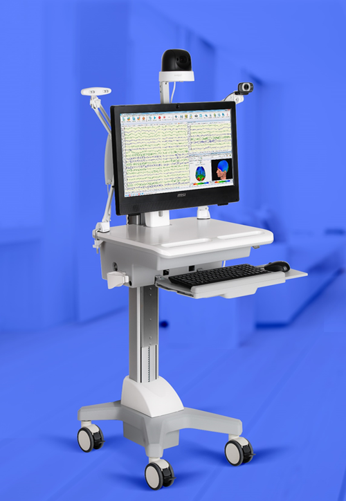
Equipment for EEG in Intensive Care Units
Registration of EEG in intensive care units imposes additional restrictions on the characteristics of the equipment. A mobile complex based on a modern electroencephalograph with a reliable noise reduction system and the highest signal recording quality is needed.
Video monitoring and tests with stimulation is a mandatory requirement for such a complex.
It is not uncommon for EEG registration in intensive care units not from scalp electrodes, but from invasive electrodes installed directly on the cerebral cortex.
The number of registration channels can be from 16 up to 64.
EEG in Intensive Care Units
Here is few examples of EEG systems in ICU:
Quantitative EEG in Intensive Care Units
Among the software requirements, in addition to convenient means of viewing the recorded EEG, there are various kinds of trends such as:
- Amplitude-integrated EEG
- The trend of the frequency spectrum (DSA)
- Alpha-rhythm variability trend
- EEG amplitude average trend
- Trends of wave rhythm indices.
Remote access to the EEG data, seizure detector, burst-suppression detector, warnings notifications and other software functions are useful in practise.
Diagnosis of brain death
There are several methods to confirm the diagnosis of brain death. One of the most common is the EEG examination. In accordance with the requirements of national laws and in accordance with international recommendations for the diagnosis of brain death, it is necessary to record EEG on at least 16 channels for at least 30 minutes. The amplitude of the EEG signal during this time should not exceed 5 μV. It is clear that in order to implement these requirements, the level of internal noise of the amplifier should be significantly less than 5 μV.
Another important requirement is confirmation of the absence of responses when registering auditory or somatosensory EPs. That is, in addition to EEG, it is also necessary to register EP and make sure that there is no brain activity in response to stimulation of different modality (auditory, current, flash, pain).
Solution for diagnosis of brain death
- Registration up to 23 EEG channels
- Low level of internal noise
- Automatic measurement of the amplitude of the recorded EEG signal
- The presence of built-in stimulators (photo, audio, current, pattern)
- Possibility of recording long and short latency evoked potentials of all modalities
- Possibility of mobile performance with a laptop
- Possibility of use as a 4-channel electroneuromyograph
MONITORING THE DEPTH OF ANESTHESIA UNDER THE CONDITIONS OF A MEDICAL SEDATION
Sedation is a controlled level of drug-induced depression of consciousness, at which protective reflexes are preserved, adequate breathing is provided, and there are responses to physical stimuli or verbal commands. Sedation is also defined as a complex of medication and non-medication, designed to provide the patient's physical and mental comfort and facilitate the technique of nursing in the department of anesthesiology, resuscitation and intensive care.
- Excitement is quite common among ICU patients: in 16-71% of cases, with pronounced agitation in 16-46% of cases.
- Insufficient or excessive sedation leads to post-traumatic stress disorder, observed in 15-27% of resuscitation patients, significantly impairing their quality of life.
Currently, several different techniques have been developed for the objective control of sedation. One of the simplest and at the same time reliable ways to control the depth of anesthesia is to register the EEG in just one channel.
Intravenous and inhalation anesthetics have different effects on the EEG, and their equipotential concentrations produce very different EEG frequencies.
Nevertheless, the general rule for changing the EEG pattern under their sedation, proposed by Faulconer A.J. and Bickford R.G. (1990), which manifests itself as "a slowdown in frequency and an initial rise followed by a decrease in EEG amplitude depending on the clinical range of anesthesia depth“, can be applied to most anesthetics in use today.
Solution for monitoring depth of anesthesia
- High accuracy in measuring the depth of anesthesia
- Easy to use
- Has built-in artifact detection algorithms
- Proven Efficiency
CONCLUSIONS:
EEG in ICU is a useful tool for monitoring the functional state of the central nervous system.
In cases discrepancy between the duration of impaired consciousness and the severity of the underlying disease or discrepancy between the level of depression of consciousness and the severity of the course of the disease, EEG in ICU can help to understand functional brain state and status, make timely correction of treatment and and make disease outcome prognosis:
Also EEG in ICU can be used for brain death diagnosis and anesthesia depth monitoring.