Electroencephalography (EEG) - sensitive non-invasive method for studying the functional state of the brain.
Method History
The beginning of the study of the electrical processes of the brain was laid by D. Raymond (Du Bois Reymond) in 1849, which showed that the brain, like the nerve and muscle, has electronic properties.
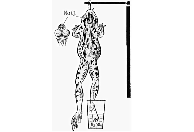
In 1882, I.M.Sechenovpublished the work "Galvanic phenomena on the medulla oblongata of the frog", in which the fact of the presence of rhythmic electrical activity of the brain was first established.
The beginning of electroencephalographic research was laid by V.V. Pravdich-Neminsky, having published in 1913 the first electroencephalogram recorded from the dog’s brain.
The first human EEG record was obtained by the German psychiatrist Hans Berger in 1928. He also suggested recording the biocurrents of the brain as an “electroencephalogram”.
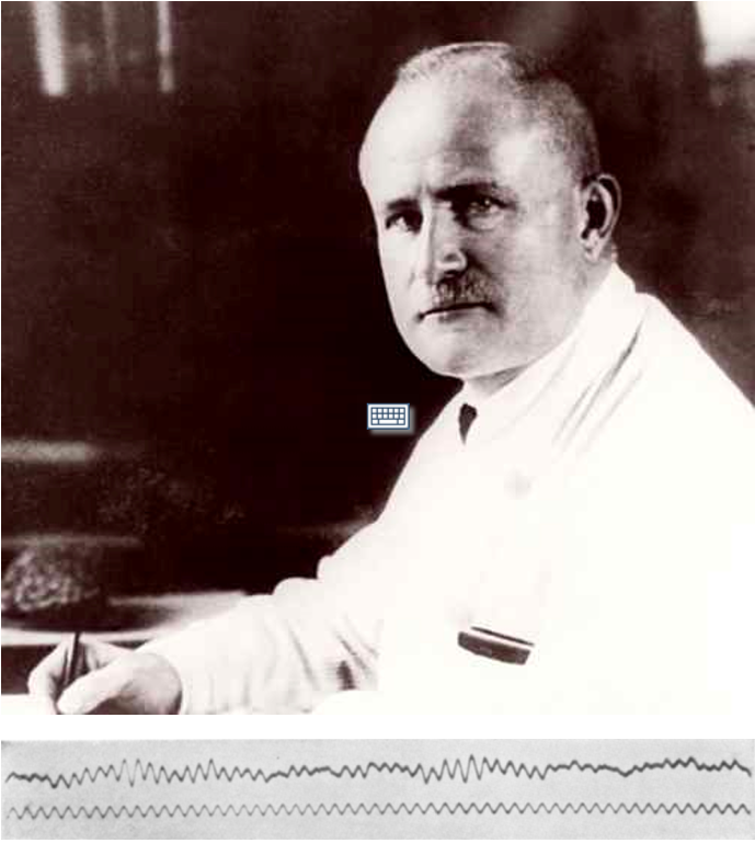
The result of Berger’s work was the article “On the Human EEG” (1929), which described the basic EEG semiotics of a healthy person.
By the mid-1950s, almost all of the currently known EEG semiotics was accumulated and systematized. The main hypotheses of generating the total bioelectric activity of the brain were formed.
In the 1990s, computer methods for processing, visualizing and analyzing EEGs were born and began to flourish, significant progress was made in the technique of recording EEGs. To register an EEG, shielded cameras are no longer required, you can conduct an examination right at the patient's bedside.
Currently, EEG is a common available method for studying the functional state of the brain, is a reliable tool in the hands of a neurophysiologist, neurologist, epileptologist, and specialist in perinatal medicine in the diagnosis of diseases such as:
- epilepsy,
- encephalopathy,
- Alzheimer's disease,
- brain tumors,
- head injury,
- sleep disturbances,
- mental disorders.
The possibilities of EEG are not yet fully exhausted. Promising scientific developments are being carried out in the field of constructing brain-computer systems, which will allow the thought to control computerized equipment. Innovative rehabilitation systems for stroke patients have already been applied using EEG-based biofeedback trainings. In the future, scientists hope to learn to recognize speech and even human thoughts using microscopic EEG electrodes implanted in the brain.
Modern EEG technique
Currently, for registration of the EEG, mainly only digital computer systems are used, which allow not only high-quality registration of the EEG signal, but also view the recorded data in various montages, with different scales, filters, quickly and accurately measure the amplitude and frequency of the EEG curves .
Modern computer programs for the mathematical processing of EEGs help to analyze the recorded signal and prepare a examination conclusion in a fully automatic mode.
The principle of a modern encephalograph
A modern EEG recorder is an electronic unit consisting of two main elements: an amplifier and an analog-to-digital converter (ADC). In the end, the technical characteristics and capabilities of the recorder add up to the parameters of these two components. Low-amplitude electrical activity of the brain is recorded by electrodes installed on the head of the subject, wired to the inputs of the recorder amplifiers, amplified many times, digitized by the ADC and transmitted to the computer, where it is already filtered, displayed on the monitor screen, subjected to mathematical processing at the request of the doctor.
Electrodes for EEG acquisition
To register an EEG, there are several types of different electrodes, which can be divided into two types:
- Non-invasive EEG electrodes (superimposed on the scalp).
- Invasive EEG electrodes (superimposed on the open brain).
In most cases, scalp non-invasive electrodes are used to record the EEG. These are, as a rule, bridge, cup electrodes or electrode systems with pre-installed electrodes. For better electrical contact with the scalp when applying EEG electrodes, saline or electrode gel is used.
There are so-called “dry” EEG electrodes that do not require the use of a gel.
The most modern can be considered active electrodes, which in their case already contain an ADC and an EEG signal amplifier. Using these electrodes, you can get the highest quality EEG signal.
EEG montages
EEG electrodes on the head of the subject are placed in strictly defined places according to the international system “10-20%” or “10-10%”. During the examination, the difference in electrical potentials between the electrodes on the head and the so-called reference electrode is continuously recorded, which can be applied, for example, to the earlobe, nose or forehead. The totality of all EEG electrodes superimposed on a patient is commonly called registration montage. Monopolar and bipolar montages are divided. In monopolar montage, several leads are recorded relative to the common reference electrode. In bipolar - each derivation has its own referent.
EEG rhythms
During the registration of the EEG examination, the electrical activity of the brain under each electrode is recorded. This activity is presented along the time axis in the form of a trace. In this trace, it is customary to distinguish waves of certain frequency ranges, which are called EEG rhythms. The main EEG rhythm of a healthy person in a state of relaxed wakefulness is considered the alpha rhythm (8-14 Hz). When solving, for example, mathematical tasks, the brain to a greater extent begins to generate a beta rhythm (14-35 Hz), and in a person’s dream, delta (0.5 - 4 Hz) and theta (4-8 Hz) rhythms dominate. By the presence or absence of certain rhythms, depending on the condition of the patient, according to their localization, the doctor can make a conclusion about the functional state of the brain of the subject.
Stationary EEG machine
Modern computer systems for EEG acquisition are divided into stationary and portable.
Stationary systems are used to record routine EEG examinations and long-term EEG video monitoring.
Thanks to their autonomy and wireless interface, portable systems can also be used for EEG registration even at the patient’s home.
Portable EEG recorder
The advantages of portable EEG systems are that they are miniature, can be placed directly on the body of the patient and, thanks to the presence of a wireless interface, allow the patient to move freely during the examination.
Example of EEG traces
Modern computer systems, in addition to directly recording and reviewing EEG traces, allow you to simultaneously review synchronous video and the results of mathematical analysis of recorded data in the form of trends, tables, topographic maps, histograms, etc.
EEG functional tests
During the EEG examination, EEG is usually recorded in various conditions of the patient. So, a typical routine EEG examination in accordance with different international recommendations should include the following functional tests:
- Background test - carried out with eyes closed in a state of calm wakefulness. During this test, an alpha rhythm is recorded in a healthy person (sample duration 20-30 minutes).
- Eyes opening - during this test, usually the intensity of the alpha rhythm drops sharply (depression of the alpha rhythm) (duration 20-30 sec.).
- Eyes closing - alpha rhythm is restored (duration 20-30 sec.).
- Flash stimulation - a provocative test is carried out with a different frequency of presentation of light flashes to the patient (duration 3-5 minutes). In patients with epilepsy, a series of flashes can cause a seizure. In healthy people, the assimilation of the rhythm of stimulation is recorded.
- After flash stimulation, it is carried out to check the restoration of background EEG activity (duration 2-3 minutes).
- Hyperventilation - a provocative test with deep breathing of the patient (duration 3 minutes). Deep breathing can provoke the appearance of epileptiformactivity on the EEG of sick people.
- After hyperventilation, it is carried out to check the restoration of background EEG activity (duration 1-2 minutes).
EEG filters
According to EEG rhythms traditional frequency band for EEG signal acquisition is 0.5–35Hz. Such filters allow to remove most of artefacts and record clear EEG activity of the brain.
But according to IFCN recommendations for modern EEG systems it is recommended 0.5–70Hz band to avoid missing of epileptiformactivity on EEG.
According to this new recommendation, minimum sampling rate for EEG recording should be at least 200 Hz.
EEG artifacts
Modern electroencephalographs have high sensitivity and good noise immunity, which allows you to record high-quality EEG in almost any conditions. However, registration of spurious signals or noise is still possible, since any EEG complex ultimately registers electrical activity, and the electrical activity of the brain is much lower than, for example, the heart and muscles. There are several types of typical artifacts when registering an EEG:
Physiological:
- ECG artifact
- EOG artifact
- EMG artifact
- GSR artifact
Non-physiological:
- Electrode movement artifact
- Electrical artifact from the mains
Modern software packages for EEG processing contain various tools for the extraction and even removal of artifacts from the EEG, but nevertheless, the doctor who analyzes the EEG data should be able to isolate all types of artifacts and clearly separate them from the true EEG.
EEG mathematical analysis
With the advent of the first computerized complexes for EEG registration, various mathematical methods of processing and analysis began to develop rapidly, which significantly expanded the diagnostic capabilities of the method. In addition to methods for suppressing artifacts and automatic calculation of rhythm amplitudes, modern EEG complexes, as a rule, contain the following types of mathematical processing of EEG:
- Amplitude analysis.
- Spectral analysis.
- Correlation analysis.
- Coherent analysis.
- Periodometrical analysis.
- Analysis of independent components.
The results of the analysis can be presented in the form of tables, graphs, topographic maps.
During the registration of the EEG, a separate trace is recorded from each electrode. Knowing the location of the electrode and the totality of electrical signals from all the electrodes, a computer program can build a model of the electrical activity of the brain and display it on the monitor screen in the form of 2D and 3D topographic maps. In addition, using various mathematical models of the distribution of electrical signals in the brain, it is possible to determine the localization of the sources of pathological activity and see them on a three-dimensional model.
EEG guidelines and clinical recommendations
- National recommendations and guidelines.
EEG techniques overview
The range of application of EEG in clinical practice is currently quite wide. The list of commonly used techniques includes:
- Routine EEG examinations.
- Long-term EEG video monitoring.
- Evoked potentials of the brain.
- Cerebral function monitoring.
- EEG in intensive care units.
- Invasive EEG examinations.
- Monitoring Depth of Anesthesia.
- Diagnosis of brain death.
- Polysomnography.
- Therapy with biofeedback.
- EEG + TMS.
- Scientific researches.
Routine EEG examinations
For routine EEG examinations, a stationary (rarely mobile) complex is usually used. As a rule, bridge or cup EEG electrodes (less cap, needle electrodes) are applied, in accordance with the international system “10-20%” or “10-10%”. Typically, recording is carried out in monopolarmontages with the ability to switch to bipolar montages. A minimum of 19 EEG derivations are recorded. It is also recommended to register one ECG channel and two EOGchannels.
The duration of the examination is about 20-30 minutes, depending on the purpose of the examination and ongoing functional tests.
Typical functional tests: background recording, opening / closing eyes, flash stimulation, hyperventilation.
At the end of the examination, a report is formed with a conclusion on the results of the examination and the condition of the patient.
Neurosoft solution for routine EEG
«Neuron-Spectrum-63» in a delivery set for routine EEG examinations :
- 19-21 channel for registration of EEG
- Dedicated ECG and EOG channels
- Compliance with IFCN recommendations for routine EEG examinations
- Impedance Indication on Electrode Connectors
- Possibility of using various electrodes and electrode systems
- High quality registration in any unscreened room
- Flexible software
Long-term EEG video monitoring
Long-term EEG video monitoring can be carried out both in a hospital (in a specially equipped room) and at the patient’s home (when using portable EEG recorders). EEG from 21 to 64 EEG electrodes is recorded. Typically, cup electrodes or electrode systems are used.
Synchronized with EEG high-resolution video is recorded to compare the clinical picture with the electrical activity of the brain. In modern video monitoring systems, as a rule, network video cameras are used.
The duration of the examination can be from several hours to several days. As a rule, the purpose of such examinations is to confirm the diagnosis of epilepsy or to clarify the focus of paroxysmal activity, so the software of such systems should search for and highlight the phenomena of epileptiform activity on the EEG.
Here is one of the first video EEG complex with mirrors system:
And here is modern video EEG laboratory:
Often, the operator uses a dual-monitor mode to view the monitoring results.
Neurosoft solution for long-term EEG video monitoring
«Neuron-Spectrum-64» in delivery set «Video»:
- 21–25 EEG channels
- High quality continuous synchronous EEG and video recording
- Support for up to 3 network cameras with Full HD resolution
- Possibility of using various electrode systems
- Online impedance measurement
- Ability to register with invasive electrodes
- Stationary or mobile design
- Automatic search and extraction of spikes, sharp waves and other types of epileptiform activity on the EEG.
Ambulatory long-term EEG video monitoring
With the advent of portable EEG recorders, EEG video monitoring has become possible not only in the hospital, but also at the patient’s home. This is more convenient for the patient, cheaper for the medical clinic, and moreover, the effectiveness of such an examination, as a rule, is higher, since in the usual conditions of life for the patient, epileptic seizures occur more often.
For registration, a portable EEG recorder with the ability to record from 19 to 32 EEG channels is required.
The duration of the examination can be up to 3 days, while the EEG machine should provide continuous recording of EEG and video around the clock.
For video monitoring, from 1 to 3 wireless video cameras can be used. Sometimes video recording is used when recording motion only.
Modern systems allow the operator to remotely view the recorded data in real time via the Internet and, if necessary, instruct the patient.
Neurosoft solution for ambulatory long-term EEG video monitoring
«Neuron-Spectrum-AM» in delivery set for «Ambulatory EEG video monitoring»:
- Fully autonomous portable EEG recorder
- Registration of 21 EEG channels
- High-quality long-term synchronous registration of EEG and video at the patient’s home (up to several days)
- Mobile wireless video cameras with Integrated microphone and day/night mode
- Freedom of movement of the patient in familiar conditions
- Possibility of remote monitoring of the examination (via the Internet)
Evoked potentials of the brain
To acquire the evoked potentials of the brain, the EEG is averaged in response to a photo, audio, current, or pattern stimulation. With the help of electroencephalographs, as a rule, long-latency EPs are recorded.
Common cognitive EP paradigms:
- P300, MMN, CNV
- GoNoGo
- P50
- N400
- OddBall
- TOVA (Test of Variables of Аttention)
- StroopTask
- VCPT (Visual Continuous Performancе Task)
To register the EP, equipment with a low level of internal noise, high sampling rate, and accurate synchronization with the stimulator is required.
Neurosoft solution for evoked potentials
«Neuron-Spectrum-4/EPM» in delivery set «Long-latency EP»:
- Registration of long-latency EPs of any modalities on the 21 EEG channel
- Low noise and high sampling rate
- Built-in photo, audio, current and pattern stimulator
- Ability to work with external stimulators
- Expandable to a fully functional four-channel electroneuromyography
Cerebral function monitoring (CFM)
Cerebral function monitoring is indicated for newborns born prematurely, with deviations or suspicions of defects in the development of the central nervous system. The rules for monitoring newborns are described in the “Atlas of AMPLITUDE-INTEGRATED EEGs in the NEWBORN”[14] and in “The American Clinical Neurophysiology Society’s Guideline on Continuous Electroencephalography Monitoring in Neonates” [13].
In accordance with international recommendations for cerebral function monitoring, continuous recording of EEG from needle, cup or disposable hydrogel electrodes in one or two derivations with an interelectrode distance of more than 75 mm is used. For analysis, the amplitude-integrated EEG (aEEG) trend is used, which has characteristic patterns typical of various kinds of deviations in the development of the central nervous system.
Computer systems for cerebral function monitoring are usually built on the basis of a two-channels electroencephalograph and are located on a mobile stand/cart.
For ease of use in intensive care units, such devices usually use a computer with a touch screen.
Also, monitoring complexes are usually equipped with video cameras to determine the moments of feeding the baby and identify other recording artifacts.
For ease of applying electrodes, some complexes are equipped with special remote units.
Such system in use:
The software of such complexes should provide viewing of aEEG trends with the possibility of automatic or manual interpretation of patterns, native EEG traces and video.
In addition, in the program, the operator can set labels and markers of events, view the results of EEG analysis and prepare examination reports.
The software of modern monitors supports the distribution of alarm notifications on the progress of the examination and the ability to remotely view the progress of the examination on a local network or via the Internet.
Neurosoftsolution for CFM
«Neuromonitor»:
- Up to 11 reference EEG channels
- Built-in neuromonitoring montages
- The portable patient unit
- All-In-One computer with touch screen
- Video registration
- Automatic or manual interpretation of aEEG patterns
- Notifications about aEEG patterns changing or epilepsy form activity detected
- Remote monitoring of several examinations
For use in ICU, the complex can be equipped with 16, 19, 21 or 32-channel electroencephalograph.
Using the observation post option, one operator can simultaneously monitor the progress of several examinations. Also, a report on the progress of each examination can be sent automatically with a specified period of time to the specified email address.
EEG in Intensive Care Units
Registration of EEG in intensive care units imposes additional restrictions on the characteristics of the equipment. A mobile complex based on a modern electroencephalograph with a reliable noise reduction system and the highest signal recording quality is needed.
Video monitoring and tests with stimulation is a mandatory requirement for such a complex.
It is not uncommon for EEG registration in intensive care units not from scalp electrodes, but from invasive electrodes installed directly on the cerebral cortex.
The number of registration channels can be from 19 upto 64.
Among the software requirements, in addition to convenient means of viewing the recorded EEG, there are various kinds of trends such as:
- Amplitude-integrated EEG
- The trend of the frequency spectrum (DSA)
- Alpha-rhythm variability trend
- EEG amplitude average trend
- Trends of wave rhythm indices.
Invasive EEG examinations
Sometimes registration of EEG from surface (scalp) electrodes is not enough. For example, to accurately localize the focus of epileptiform activity before surgical treatment of epilepsy, when mapping the motor, sensory or speech areas of the cerebral cortex during an operation to resect tumors and neoplasms in the brain.
There are several types of invasive EEG electrodes:
- Cortical / subdural electrodes (strips / grids)
- Needle electrodes (stereo EEG)
- Sphenoidal electrodes
For registration of EEG from invasive electrodes, special conditions are imposed on the equipment, for example, increased requirements. A large number of recording channels, a high quantization frequency, a wide frequency range of the amplifier, and a low level of intrinsic noise are required.
The number of invasive electrodes within one examination can reach 64 or more. Frequently, high-frequency oscillations are also recorded from invasive electrodes, the amplitude of which is small, and the frequency range can reach 500 Hz. Not every electroencephalograph can cope with high-quality registration of such a signal.
High Frequency Oscillations (HFOs):
- 80-500 Hz,
- up to 250 Hz - ripples,
- > 250 Hz - fast ripples.
Neurosoft solution for invasive EEG
Neuro-Spectrum-65 in delivery set «Mobile EEG video monitoring»:
- Registration from 39 to 78 EEG channels
- Low level of noise
- High sampling rate
- Wide input range of the amplifier in frequency and amplitude
- Mobile complex convenient to use both in the ward and in the operating room
- High resolution video monitoring
- Possibilities of 2D and 3D topographic mapping of the brain
Diagnosis of brain death
There are several methods to confirm the diagnosis of brain death. One of the most common is the EEG examination. In accordance with the requirements of national laws and in accordance with international recommendations for the diagnosis of brain death, it is necessary to record EEG on at least 16 channels for at least 30 minutes. The amplitude of the EEG signal during this time should not exceed 5 μV. It is clear that in order to implement these requirements, the level of internal noise of the amplifier should be significantly less than 5 μV. Another important requirement is confirmation of the absence of responses when registering auditory or somatosensory EPs. That is, in addition to EEG, it is also necessary to register EP and make sure that there is no brain activity in response to stimulation of different modality (auditory, current, flash, pain).
Neurosoft solution for diagnosis of brain death
- Registration up to 23 EEG channels
- Low level of internal noise
- Automatic measurement of the amplitude of the recorded EEG signal
- The presence of built-in stimulators (photo, audio, current, pattern)
- Possibility of recording long and short latency evoked potentials of all modalities
- Possibility of mobile performance with a laptop
- Possibility of use as a 4-channel electroneuromyograph
Monitoring Depth of Anesthesia
Another important task that can be solved using EEG is monitoring the depth of anesthesia.
During the operation, it is very important for the anesthetist to have objective tools to control the patient's sedation level. Both too large and too small doses of anesthesia are equally dangerous.
Currently, several different techniques have been developed for the objective control of sedation.
One of the simplest and at the same time reliable ways to control the depth of anesthesia is to register the EEG in just one channel.
Neurosoft solution for monitoring depth of anesthesia
- High accuracy in measuring the depth of anesthesia
- Easy to use
- Has built-in artifact detection algorithms
- Proven Efficiency
Polysomnography (PSG)
We often forget that we spend a third of our life in a sleep. Modern medicine knows many different sleep disorders that significantly reduce the quality of life and the level of health. Insomnia, parasomnia, obstructive apnea syndrome - these are just some of them. For the diagnosis of sleep disorders are special medical devices was developed: polysomnographs. The AASM single out the following types of such devices:
- Type I - stationary PSG recorders operating under the supervision of medical personnel
- Type II - portable PSG recorders operating without the supervision of medical personnel
- Type III - devices for cardiorespiratory monitoring
- Type IV - devices for detecting signs of sleep apnea
The AASM Manual for the Scoring of Sleep and Associated events, 2018 [12].
AASM classifications for PSG devices:
Polysomnography in sleep lab:
PSG electrodes and sensors:
Software for sleep scoring:
Neurosoft PSG solutions
- Stationary polysomnograph (Type I)
- A complete set of PSG channels in accordance with the recommendations of the AASM
- Ability to use a patient unit
- Possibility of synchronous video recording of PSG examination
- Portable expert class polysomnograph (Type I, II)
- A complete set of PSG channels in accordance with the recommendations of the AASM
- Wireless interface for transferring data to a computer
- Built-in memory card for storing PSG data
- Type III portable cardiorespiratory monitor
- Simplicity and usability
Therapy with biofeedback (BFB)
The principle of the biofeedback technique is that the patient is given feedback about the physiological processes taking place in his body. Feedback can be a game, for example, imagine a computer game in which a boat floats at a speed that directly depends on the speed of your heartbeat. Similar types of feedback exist for EEG signals. For example, indicators such as amplitude, frequency, and wave rhythm index can be taken as the basis for biocontrol. The scope of the EEG-based biofeedback technique is constantly expanding. With the help of a series of sessions of such trainings lasting 30-40 minutes a day, a child, for example, can cope with hyperactivity (Attention deficit hyperactivity disorder (ADHD)). Also, BFB trainings are used in the fight against alcoholism, smoking.
Recently, more often BFB -therapy has been used in the rehabilitation of stroke patients.
BFB training usually requires a two-monitor stationary system based on an electroencephalograph or a polygraph with the ability to output feedback to the patient’s monitor in the form of video, audio, or game information.
Neurosoft solution for biofeedback
- Dual monitor stationary system
- Ability to provide feedback on parameters: EEG / EMG / ECG / respiration / SpO2 / body temperature
- Visual, sound, game feedback
- Flexibility to configure and manage training templates
- Training success trend
EEG + TMS
Currently, solutions for recording EEGs during transcranial magnetic stimulation are quite exotic. Technically, the implementation of this method is fraught with many difficulties, since at the time of applying a magnetic stimulus to the EEG amplifier, a huge artifact signal arrives that must be ignored. Only some modern electroencephalographs have a combination of technical parameters that allow them to be used for this technique.
Nevertheless, the prospects for the development of the EEG + TMS technique are quite broad. First of all, it is currently used to monitor the recovery process of the patient’s brain after a stroke. EEG + TMS is also used in numerous scientific studies and most likely in the near future this technique will expand its field of application and become more popular.
Neurosoft solution for EEG + TMS
Neuron-Spectrum-6G + Neuro-MS:
- Due to their outstanding technical characteristics, the modern devices of the Neuron-Spectrum-6G series are one of the few electroencephalographs that can be successfully used to record EEG directly during TMS and average the responses of the brain to magnetic stimuli.
- Neuro-MS magnetic stimulator has electrically silent charging mode which helps to reduce noise on EEG during recording.
Scientific researches
Despite its impressive history, the EEG method is currently a technique that has not fully revealed its potential. In attempts to unravel the secrets of human consciousness, a wide variety of scientific studies using EEG continue.
Neurorehabilitation:
The experience of many scientific studies in the field of neurorehabilitation of stroke patients has already begun to be applied in practice. With the help of complex EEG processing algorithms and feedback technology, rehabilitation sessions are conducted. The combination of this method with functional electrical stimulation can significantly reduce the duration and increase the effectiveness of the rehabilitation process.
Brain-computer interface:
Research scientists do not stop for a minute. One of the most ambitious and promising projects can be called the development of the company Neuralink led by Ilon Mask. The development team aimed at building a full-fledged brain-computer interface using microscopic minimally invasive EEG electrodes implanted into the brain using a special robot. By receiving EEG signals from the installed electrodes, scientists expect in real time to learn to recognize first simple commands, and then speech, and even human thoughts. Today it seems fantastic.
Will see…
What Neurosoft doing for EEG science?
At Neurosoft, we always remain open to innovative research, cooperate with leading world companies in the field of EEG processing, and try to implement all the most advanced technologies in our products.
For example, our programs implement online transmission of recorded EEG data using the LSL protocol (Lab Streaming Layer) for real-time processing in programs such as MathLab, Simulink, OpenVibe.
Our equipment successfully works in many research centers and in the future we plan to continue to participate in various scientific works.
List of references
- The standardized EEG electrode array of the IFCN, Clinical Neurophysiology 128 (2017) 2070–2077.
- ILAE COMMISSION REPORT, Instruction manual for the ILAE 2017 operational classification of seizure types.
- ISCEV standard for clinical visual evoked potentials: (2016 update).
- IFCN standards for digital recording of clinical EEG, Electroencephalography and clinical Neurophysiology 106 (1998) 259–261.
- American Clinical Neurophysiology Society. Guideline 8: Guidelines for Recording Clinical EEG on Digital Media.
- Diagnostic utility of invasive EEG for epilepsy surgery:
- ILAE SPECIAL REPORT. Indications, modalities, and techniques.
- ILAE CRITICAL REVIEWANDINVITED COMMENTARY. Methodology of photic stimulation revisited: Updated European algorithm for visual stimulation in the EEG laboratory, Epilepsia, 53(1):16–24, 2012.
- American Clinical Neurophysiology Society Guideline 6: Minimum Technical Standards for EEG Recording in Suspected Cerebral Death.
- AAN Clinician Guideline Supplement: Determining Brain Death in Adults, Neurology 2010.
- ACNS: Consensus Statement on Continuous EEG in Critically Ill Adults and Children, Part I: Indications, J Clin Neurophysiol2015;32: 87–95).
- The AASM Manual for the Scoring of Sleep and Associated events, 2018.
- The American Clinical Neurophysiology Society’s Guideline on Continuous Electroencephalography Monitoring in Neonates, J Clin Neurophysiol2011;28: 611–617).
- Atlas of AMPLITUDE-INTEGRATED EEGs in the NEWBORN, Second Edition, Lena Hellström-Westas, Linda S de Vries and Ingmar Rosén.