These tips will help not to make mistakes - to remove a benign node of the thyroid gland. Be guided by knowledge, and not just the fear and perseverance of surgeons who want to operate.
Eight important tips on the diagnosis of cancer in the thyroid nodes of Dr. A.V. Ushakov
1. Please note that “follicular tumor” is not a thyroid cancer (cancer is otherwise called “follicular carcinoma”).
2. Beforehand, explain to your specialist before the diagnostic procedure your requirements for the result of the study. This refers to the ultrasound data and, especially, to the “Protocol for cytological examination of the biopsy of the thyroid gland”.
3. During ultrasound, in case of nodules, a specialist should examine the regional lymph nodes of the neck. Pay attention to this.
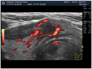
4. Together with the ultrasound protocol, at least, images of the node (or nodes) must be presented in two main projections and in the DDC (or EHF) mode. This is a modern requirement for the formation of the “Protocol of thyroid gland ultrasound” (the cost of one image is small; it does not exceed 10-15 rubles; pay attention in our film the patient holds a whole tape of images). The pictures informatively complement the characteristics of the nodal process and are therefore more important than the description alone.
5. After receiving the diagnostic "Ultrasound Protocol" carefully read it. Check the presence in it of the necessary items and their contents. Study in advance what exactly should be in the sections of the "Ultrasound Protocol".
6. Remember that "atypical cells" may be benign cells that are altered as a result of overvoltage.
7. An increase in thyroglobulin according to a blood test is not a marker of cancer. Therefore, do not worry when increasing the value of this indicator. Thyroglobulin is constantly released from the thyroid gland into the bloodstream along with hormones (T4 and T3). An increase in thyroglobulin concentration is a sign of intensive tissue activity or destruction. It can be increased with the destruction of the tissue of the benign node and with subacute thyroiditis.
8. You can always get a second professional opinion. If necessary, make a control ultrasound of the thyroid gland. It often happens that doctors with ultrasound take focal lesions for foci of nodes (in this case, a puncture biopsy is performed in vain for the patient), or benign signs are treated as malignant.