In this lesson you will learn how to see changes within the thyroid gland and understand the basic terms.
What is an ultrasound scan? Simplified is the getting and interpretation of images from the depths (organ or part of the body) using ultrasound. Ultrasound is reflected differently from body tissues, because they differ in density. This difference creates an ultrasound picture on the monitor of the ultrasound machine, which can be saved as pictures or movie-recordings.
The base image is black and white. It is called by doctors "gray scale", "gray mode" or "B-mode." Everything else is an addition (important or uninformative).
Dense fabrics look lighter (whiter). Usually, this is connective tissue. But not always a brighter image is excessively dense. Less dense areas are depicted darker, down to black, but here too - not everything is clear.
Ultrasound Terms
Ultrasound doctors most often use three basic words: isoechogenic, hypoechoic and hyperechoic. Less commonly, anechoic. You need to remember their semantic meaning.
Reference
Iso - normal, medium. Hypo is not enough. Hyper - a lot. The particle preposition "a" or "an" is denial, absence.
Echogenicity is a reflection (ultrasound from gland tissue and surrounding structures).
Isoechoic is a light gray background of varying severity. This is somewhere around 15-25% gray. Normal thyroid tissue is isoechoic.
Hypeechogenicity is a lighter background (less than 15% gray). In the thyroid gland, the connective tissue looks like this, but the tissue of the gland itself also happens, as well as calcifications or some phenomena.
Hypoechoic is a more dark gray background (more than 25% gray). Hypoechoicity is of different sizes (severity).
Anechoic - black. This is how the liquid looks (in the vessels or inside the nodes - a cyst).
See how reflection is different (i.e. echogenicity):
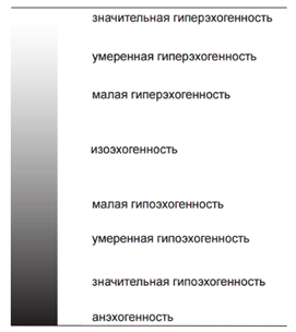
The word "echogenicity" means only reflection, and says nothing about the magnitude (quality) of such reflection!
The uzist doctor must indicate the echogenicity option.
See how the types of echogenicity are determined on an ultrasound image
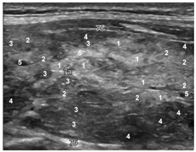
On the image:
1 - lobules with normal tissue of the thyroid gland,
2 - lobules with edema of the stroma, the initial signs of exhaustion,
3 and 4 - lobules with significantly depleted tissue and penetration of immune cells (lymphocytes),
5 – vessel
6 - connective tissue or the first signs of regeneration (there will be a lesson about this phenomenon separately).