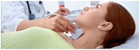
“Trust, but verify,” - this expression is also relevant to the result of ultrasound of the thyroid gland. Pay attention to the "Ultrasound Protocol" issued to you! There are certain requirements for describing important signs of change. Without this information, the diagnostic value of the “Ultrasound Protocol” will not be complete.
1. The doctor may indicate only two sizes of each lobe of the thyroid gland.
Such an error is rare. “Ultrasound Protocols”, with the indication of only two sizes of each share, were brought to our clinic from Uzbekistan, Latvia and Moldova.
The fact is, the gland lobes are quite VOLUME. These are not flat butterfly wings. Therefore, each lobe must be measured three times - in length, in width and in depth. It is not the size and size that matters. VOLUME is important. Only the volume of the thyroid gland can help the endocrinologist to determine the magnitude of the tension and exhaustion of the organ.
(Clarifying records that the thyroid volume for women is normal up to 18 ml, and for men 25 ml, do not correspond to reality and are erroneous).
In the "Ultrasound Protocol" should be indicated three sizes of each share, the volume of shares and the sum of their volumes.
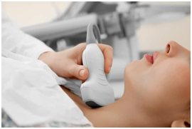
Such an ultrasound probe does not examine the thyroid gland. This convex (arcuately convex) sensor for abdominal organs. The head is located almost correctly (not bent forward).
2. Diffuse changes.
Very often, ultrasound doctors (sonologists) write in the DESCRIPTION section that some “heterogeneous” (ie heterogeneous) “hypoechoic” (i.e. dark) diffuse (i.e. common, non-nodal) changes are revealed and without indicating their QUANTITY, location in the gland and morphological essence, they also repeat in CONCLUSION that there are "diffuse changes" and they correspond to AIT.
What does all this mean? Everything is simple. The doctor revealed some changes that do not correspond to the nodes, and wrote so - these are some non-node changes (i.e. diffuse changes). What are these diffuse changes?
In the Thyroid Clinic, Dr. A.V. Ushakov distinguish 11 morphological variants of the diffuse process in the thyroid gland.
In fact, ultrasound doctors do not know the true structure of the thyroid gland. They do not know:
* the real size of the lobules and follicles,
* natural segmental organization of glandular lobes,
* neurovascular structure of gland segments.
Therefore, empirical attributes are used to describe the diffuse process or simply name it, but do not describe it.
3. How much high-grade tissue?!
An ultrasound doctor should indicate the amount of tissue that looks macrostructurally complete (in percent for each lobe).
This tissue (isoechogenic and slightly hypoechoic) is a source of hormones. If there is a lot of such tissue, and a blood test shows an increase in TSH with normal amounts of thyroid hormones (T3fr. And T4fr.), then you do not need to introduce additional hormones with the drug. Moreover, in this case, an excessive dose of a hormonal medication is not needed (after all, endocrinologists like to immediately prescribe 50 mcg - the average dose).
Unfortunately, none of the sonologists indicates this important circumstance in the “Protocol of ultrasound”.
4. Vascularization.
You are a vessel. Vascularization is vascularity. So they write most ultrasound doctors. But often, doctors do not report anything at all about vessels and blood flow in the thyroid gland.
If you do not see the words “vascularization”, “blood flow”, the abbreviations CDK, EDK (ED) and commentary on them, then the most important part of the ultrasound diagnosis of the thyroid gland is missing.
The specialist must indicate the VALUE of blood flow stress in each lobe of the gland. It is not just to report an increase, but to apply a quantitative designation - small, moderate (medium) or significant (increased blood flow).
It is also important to measure the speed of blood flow in the arteries of the thyroid gland.
What for? Then, what is the intensity of blood flow and its speed shows the magnitude of the gland overstrain.
You can learn more about Doppler ultrasound with thyroid ultrasound in our special article for patients.
5. Description of the node in the thyroid gland.
In the "Ultrasound Protocol" should be the following characteristics of the node:
• THREE node sizes (not two, not one - "diameter"; spherical nodes are a rarity) in different planes,
• location in the lobe,
• the form,
• border and contour (uniform, whether along the entire perimeter ...),
• internal structure and condition of the tissue node,
• distribution of blood flow to the node (around the perimeter, inside, ordering of blood flow),
• node blood flow intensity (without, weakened, small, moderate, significant).
In some cases, the node can be additionally described by other signs showing the presence or absence of malignancy, and the velocity of large vessels of the node is also measured (assessment of the activity of the process in the node).
The more complete the description of the “Ultrasound Protocol”, the more accurate and more useful the treatment tactics.