Let's start with a practical lesson. You will learn to see the thyroid gland on ultrasound images, understand what it should be in a healthy state, and learn the principles of ultrasound at the very beginning of diagnosis. And it's just that simple...
Before conducting an ultrasound in our Сlinic patients doubt that they will be able to understand anything. But after the ultrasound, the patient sits with me next to the ultrasound machine, then I show and explain in the pictures that I display on the screen, in what condition is his thyroid gland, how it is tense and what can be expected in the future.
Usually, after the ultrasound procedure, patients agree that they began to understand and see on the ultrasound images the thyroid gland, changes in it and normal tissue.
Ultrasound is easy
No need to be afraid not to understand. You should relax and start.
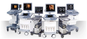
An ultrasound machine is just a computer on wheels, with a monitor and ... scanners.
The main difference is scanners. These are not flatbed office A4 paper scanners and others. For ultrasound of the thyroid gland, flat scanners are used - linear, 4 cm long (usually) or 6 cm long (less often), and several mm wide.
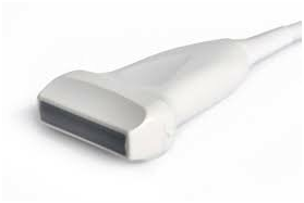
This form of the scanner allows you to penetrate ultrasound into the body and display the image on the monitor screen in the form of a flatness. The word flatness is the key!
Thyroid scan FLATNESSES for ultrasound
The thyroid gland is an organ consisting of two volume fractions that are not similar to the flat wings of a butterfly, as academicians and presenters of medical programs assure you. Therefore, each lobe (and isthmus) is studied with ultrasound in different flatnesses.
The main planes of ultrasound diagnostics are two mutually opposite - transverse (across the lobe) and longitudinal (along the lobe).Two main scanning planes for ultrasound of the thyroid gland.
Here is a TRANSVERSE projection of the left lobe of the thyroid gland (from my book on ultrasound for specialists). The thyroid gland is indicated by number 1. The healthy gland looks light gray almost homogeneous into a small gray speck.
Here's another ultrasound scan of a healthy thyroid gland from another patient. (Pay attention to the lower right corner, where the schematic image of the thyroid gland is located, where the location of the ultrasound sensor scanner is shown in the projection of the left lobe).
In a longitudinal projection, the same healthy left lobe of the thyroid gland looks like this (in the "trapezoid" or "convex" mode):
How does an ultrasound of the thyroid gland begin?
1. The doctor places a sensor scanner over one of the thyroid glands and conducts in a transverse projection above it from the “upper” pole to the “lower” lobe, while examining the image of the gland on the screen. This consecutive conducting over a share allows to see all transverse projections and to make out all local changes in a share.
2. Then the doctor changes the position of the sensor scanner to a longitudinal one and also studies the structure of the lobe, moving the sensor to the sides and also examining the lobe, but in the longitudinal direction.
After examining the condition of the lobe of the gland, the doctor measures the proportion. About this and features related to size and more, there will be the next lesson on ultrasound of the thyroid gland.